Occasionally, babies are born with one of many different forms of congenital heart disease where there is only one normally functioning ventricle. This single ventricle has to perform the work of two ventricles, pumping blood to both the lungs (pulmonary circulation) and the rest of the body (systemic circulation).
This extra load on one ventricle and mixing of what should be separate circulations is repaired through a series of operations culminating in the Fontan procedure. After repair, the pulmonary and systemic circulations are separated. The single ventricle does the hard work of pumping blood into the systemic circulation while blood flow is provided to the lungs without having to go through the ventricle.

The creation of this “Fontan circulation” is a lifesaving procedure, though it does require careful monitoring to ensure the ventricle is functioning properly and adequate flow is being provided to the lungs and the rest of the body.
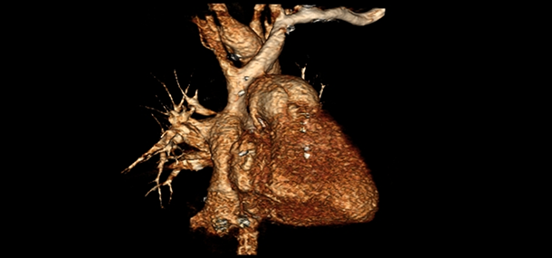
Imaging provides valuable information at different points between the stages of surgical repair as well as later on in life to ensure the Fontan circulation is functioning adequately. Fontan patients are frequently imaged with echocardiography (cardiac echo). Sometimes, a cardiac echo cannot provide answers to all the questions. In those cases, a cardiac MRI may be requested. Cardiac MRI is used to measure cardiac chamber sizes, ventricular function and look for complications associated with the Fontan circulation. In addition, sometimes the images from a cardiac MRI or CT are used to make a life-size model with a 3D printer. The model aids surgeons in planning for surgery and looking at the relationships between different parts of the heart.

The Fontan circulation doesn’t just affect the heart. The high venous pressure required to get blood into the lungs without going through a pumping ventricle affects other organs in the body. Liver congestion occurs in nearly all Fontan patients. Sometimes, the liver congestion can progress to chronic liver disease, which can result in additional complications. The liver is often monitored with ultrasound, computed tomography (CT or CAT scan), or MRI.
Elastography is a relatively new technique that determines how stiff a particular tissue is by measuring how fast mechanical waves move through the organ. Liver congestion and most forms of chronic liver disease cause the liver to become stiffer, which can be measured by elastography and monitored over time to look for progressive liver disease. Cincinnati Children’s is one of the few hospitals in the nation that routinely uses MRI elastography to monitor for liver disease. It is with this partnership of surgery and imaging that we are constantly striving for better ways to care for you and your children.
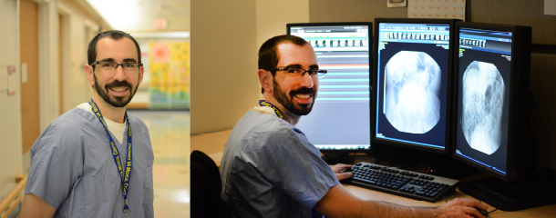
Contributed by Dr. Daniel B. Wallihan and edited by Tony Dandino (MR Technologist).
Tony is an MRI Technologist at Cincinnati Children’s. Tony has been in his role for several years and serves as a Charge Tech, Quality Improvement Coach and Safety Coach for the MRI department. Tony has always known he wanted to work with children and in the medical field. Working at Cincinnati Children's has been the best of both worlds. Every day is something new and Tony can never wait to start the next adventure.