Most will recognize this face: Kansas City Chiefs quarterback and Super Bowl MVP Patrick Mahomes, who injured his knee in an NFL game in Oct 2019.
Dislocation is also a common complaint for a child who presents to the emergency room or a doctor’s office with a knee injury. It is typically an outside dislocation of the knee cap (patella) from its normal position in the groove at the lower end of the thigh bone (femur), and is called a patellar dislocation. [A true knee dislocation is an uncommon and more severe injury where there is extensive tearing of the major ligaments of the knee joint that results in separation of the two large bones (femur and tibia, the main bone in the calf), that form the main knee joint.]
Patellar dislocation often happens during sports or play, though it may happen spontaneously in children who have a predisposition due to anatomy and/or muscle imbalance. The patella frequently reduces on its own, though sometimes it may require medical assistance. Patrick Mahomes’ patella was reduced by a physician on the field.
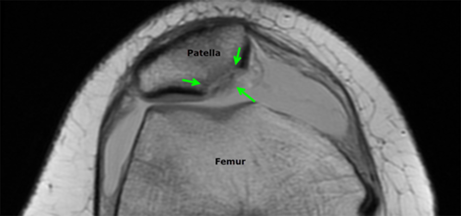
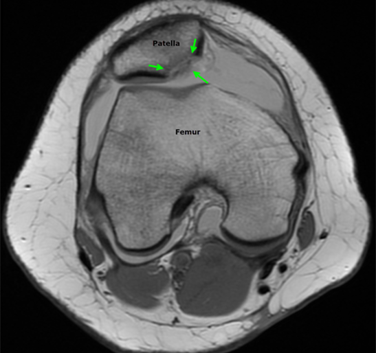
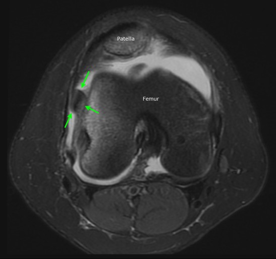
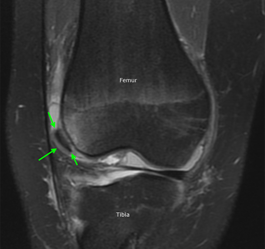
The diagnosis is typically made by history and physical examination. Knee x-rays are obtained initially to look for bone injuries, but cannot fully show the extent of soft tissue damage. For this reason, an MRI is performed soon after to assess the ligaments and cartilage and help determine whether surgery will be necessary. The MRI will also show underlying anatomical features that may predispose to the instability of the patella. If there is a significant injury to the cartilage covering the bone, a loose fragment in the joint, or tearing of the main ligament that stabilizes the patella, surgical treatment is recommended.
Luckily for Patrick Mahomes, the MRI did not show damage to the cartilage or ligaments. He was treated medically with physical therapy and able to return to play in a couple of weeks. The child with the MRI below was not as fortunate and required surgery to repair the patella. After a patellar dislocation, there is an increased risk of a recurrent dislocation, so continued follow-up and therapy are necessary.
Dr. Kathleen Emery, author; Glenn Miñano, BFA, editor; Meredith Towbin, copyeditor

Glenn Miñano is a media specialist in the Department of Radiology, providing graphic design, photography, printing, video services, and administration of the department’s online properties. His works have been published in several medical articles, such as the American Journal of Radiology and the American Institute of Ultrasound. He has been providing these services to the Radiology Department since 1996.