Prenatal imaging with ultrasound is the standard of care for all pregnancies. Sometimes, we identify abnormalities in the child before birth. One of these is known as congenital diaphragmatic hernia. When we find this abnormality, we obtain a fetal MRI to understand the anatomy and try to predict outcome.

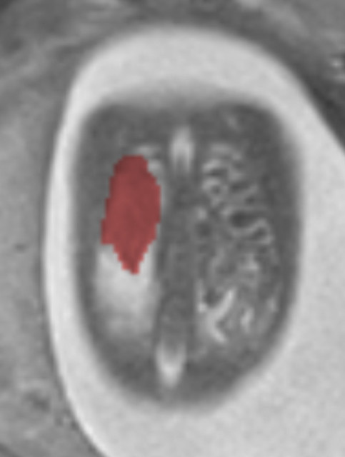
What is a diaphragmatic hernia? The diaphragm is the structure that separates the chest and the abdomen. If it does not form normally, then the bowel, stomach and liver can slide into the chest. This is what we look for when diagnosing a congenital diaphragmatic hernia. The important thing is that when these structures move into the chest, then the lungs cannot grow normally; therefore, babies with congenital diaphragmatic hernia have more difficulty breathing because the lungs are small and not normal.

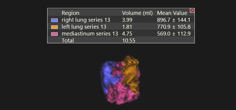
We have identified many babies with this problem. We utilize fetal MRI to calculate how much lung a baby with congenital diaphragmatic hernia has and we can compare it to normal babies at this age. With this information, with the specialist at the fetal care center, we can counsel the family and help them decide whether we should intervene in the womb or determine where the baby should be born and with what support. One of the innovative therapies for these babies whose lungs are small is to place a balloon in the airway, which creates pressure for the lungs to grow as the fluid present in the lungs cannot escape. Many times we help guide birth with extra support at Cincinnati Children’s.


With these new therapies, we are improving the outcome for babies with congenital diaphragmatic hernia at Cincinnati Children’s Medical Center.

Dr. Beth Kline-Fath, author; Glenn Miñano, BFA, editor; Meredith Towbin, copy editor
Glenn Miñano is a media specialist in the Department of Radiology, providing graphic design, photography, printing, video services, and administration of the department’s online properties. His works have been published in several medical articles, such as the American Journal of Radiology and the American Institute of Ultrasound. He has been providing these services to the Radiology Department since 1996.