Subscribe today for more stories, tips, and updates.

Cincinnati Children's Blog
  • Subscribe
  • CincinnatiChildrens.org
  • Contact Us
  • Blog
  • Healthy Living
  • Rare and Complex Conditions
  • Safety and Prevention
  • Heart Conditions
  • Learning and Growing
  • Research and Discoveries
  • ?
  • Blog
  • Healthy Living
  • Rare and Complex Conditions
  • Safety and Prevention
  • Heart Conditions
  • Learning and Growing
  • Research and Discoveries
  • ?
  • Subscribe
  • CincinnatiChildrens.org
  • Contact Us
HOME/How We Do It/Craniosynostosis: Imaging the Misshapen Head

Craniosynostosis: Imaging the Misshapen Head

Craniosynostosis: Imaging the Misshapen Head
April 9, 2019
0 Comments
By: Janet M. Adams

Is the pediatrician concerned that your baby’s head is misshapen? If so, the doctor might order a head CT to evaluate the cranial sutures to make sure that they have not closed prematurely. A baby’s head can also become misshapen from positional changes, including deformities that might have occurred in utero when the baby was developing.

The skull is composed of multiple bones that are connected at the fibrous cranial sutures. The sutures allow the skull to increase in size to accommodate brain growth throughout infancy and childhood. They also allow the skull to mold with mild overlapping of the cranial sutures during the birthing process, when a slightly smaller head size is advantageous for an easier delivery. Normally, the sutures become progressively narrowed and ossify as the child develops. The large sutures persist into early adulthood and can still be seen on radiographs and CT images as thin, irregular lines between the large skull bones.

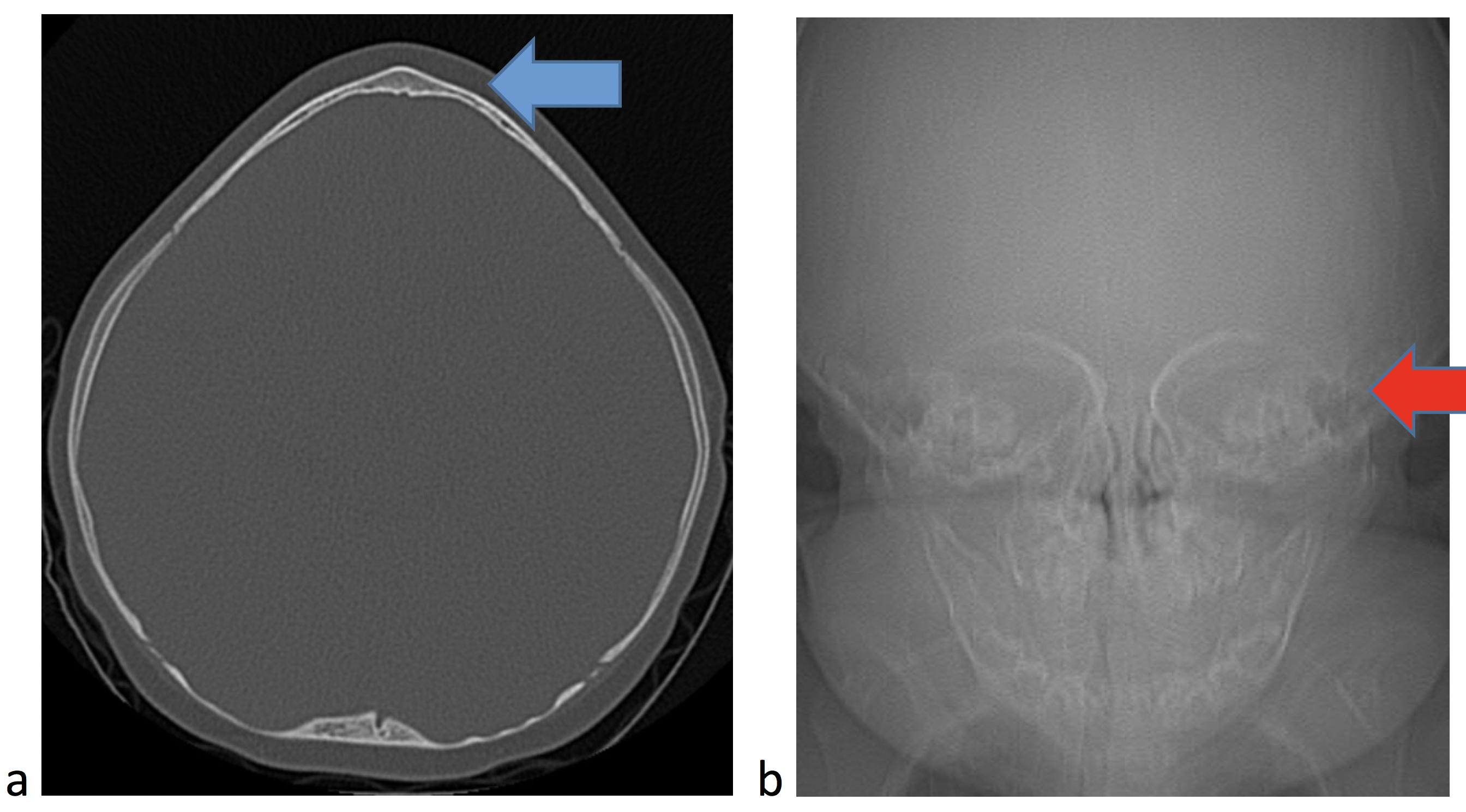
Some sutures normally close in infancy and childhood, such as the metopic suture. Even though this suture normally closes, it can also close prematurely leading to a pointed configuration of the frontal bone (Fig. 1a) and narrowing of the space between the eyes (Fig. 1b).

Fig. 1 Metopic synostosis. 1a. Axial CT with pointed frontal bone (blue arrow). 1B. Scout image shows orbits closer together than expected or hypotelorism (red arrow).
Fig. 2 Sagittal synostosis. 3D reconstruction from a head CT shows the elongated head shape as growth continues along the coronal (blue arrow) and lambdoid sutures (red arrow) in this patient with sagittal synostosis (actual suture not shown).

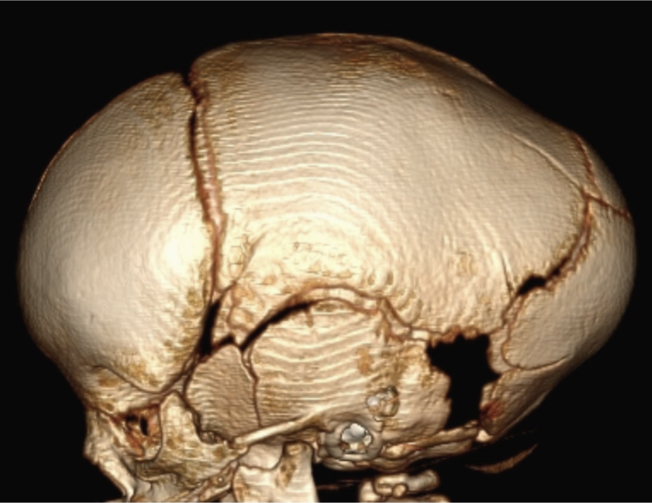
Craniosynostosis refers to the premature fusion or ossification of the cranial sutures and can occur from genetic etiologies, as well as from some metabolic disorders and mechanical changes, such as in a child with shunted hydrocephalus. With premature closure of a suture or sutures, relatively predictable head shapes and facial distortion occurs. A child with premature fusion of the sagittal suture, the most common type of craniosynostosis, presents with an elongated head shape, as the skull continues to grow along the lambdoid and coronal sutures (Fig. 2).

Fig. 3 Left coronal synostosis. Radiograph shows the typical “harlequin” configuration of the orbit adjacent to the fused left coronal suture.

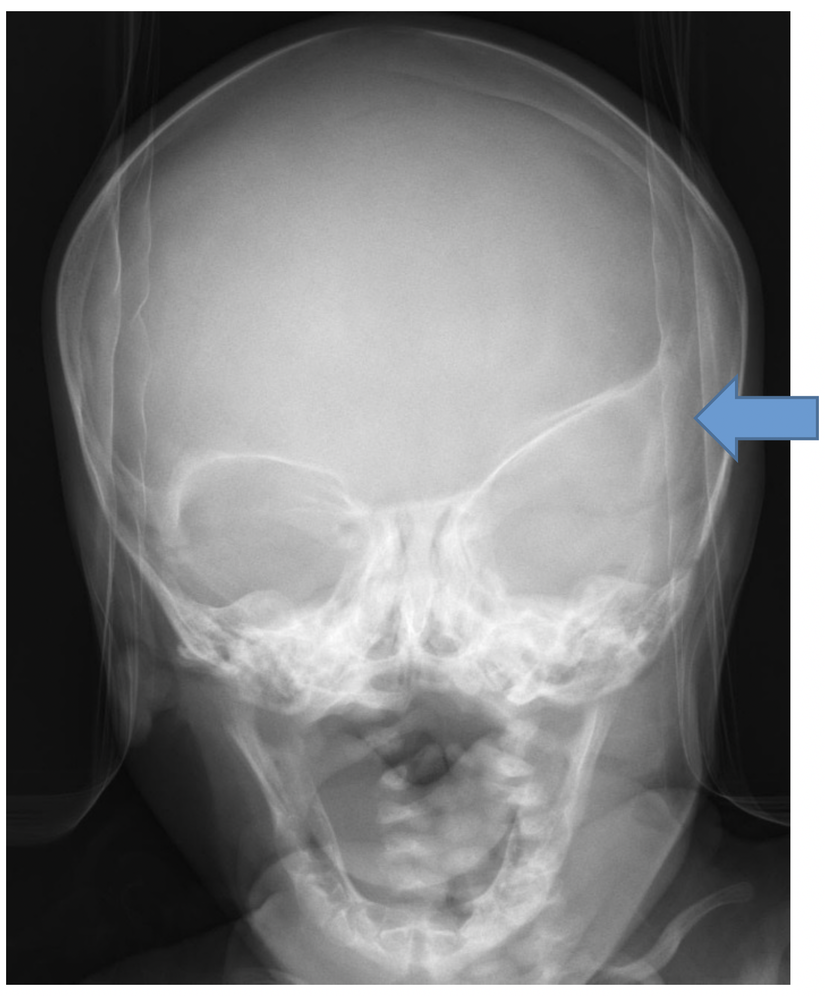
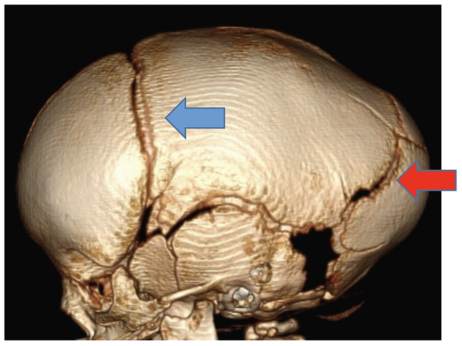
A child with unilateral coronal synostosis presents with flattening of the frontal region on the affected side, and the head shape becomes somewhat trapezoidal. On radiographs, a classic imaging finding is the “harlequin eye,” as the orbit remodels adjacent to the fused coronal suture (Fig 3).

Although radiographs and ultrasound can be used to assess the sutures, a 3D head CT readily demonstrates the normal sutures, the prematurely fused sutures, and allows the doctor to consider different treatment options. At Cincinnati Children’s, these CTs are performed with a slightly lower radiation dose, as the bones are the primary interest, and multiplanar and 3D reconstructions are computer generated without any additional imaging or radiation exposure to the infant or child.

Contributed by Dr. Marguerite M. Caré and edited by Janet Adams, RDMS, RVT, CNMT, RT(N)

Subscribe today for more stories, tips, and updates.

Related Articles

<strong>Prediction of Fontan Outcomes Using T2-Weighted MRI Radiomic Features and Machine Learning</strong> 
Prediction of Fontan Outcomes Using T2-Weighted MRI Radiomic Features and Machine Learning 
<strong>How Does Radiology Share Your Images?</strong>
How Does Radiology Share Your Images?
Radiologic Technologist Programs
<strong>MRI’s Role in Your Child’s Proton Therapy</strong> 
MRI’s Role in Your Child’s Proton Therapy 
TAGS:
  • craniosynostosis
  • CT
  • Imaging the Misshapen Head
  • radiology
Avatar photo

About the author: Janet M. Adams

Janet is a sonographer at Cincinnati Children’s. She has worked in the Ultrasound department for over 26 years, and clearly has a passion for working with children. Janet serves as a lead Safety Coach, TJC representative, and education resource for her department. She enjoys challenging exams, and is involved in local and global ultrasound research projects. When she is not at work, her 4 children and 9 grandchildren keep her very busy!

Write A Comment

Leave a Reply Cancel reply

Your email address will not be published. Required fields are marked *

No Comments Yet

Topics

  • Cancer and Blood Diseases
  • Child Development and Behavior
  • Childhood Obesity
  • Chronic Care Networks
  • Cincinnati Walks for Kids
  • Featured Faces
  • Fetal Conditions
  • Fitness and Nutrition
  • Food Allergies
  • Ghana 2012
  • Health Care Public Policy
  • Health Care Quality
  • Health Care Reform
  • Healthy Living
  • Heart Conditions
  • Hospital Operations
  • How We Do It
  • Learning and Growing
  • Meet The Team
  • Nurses Week 2012
  • Patient Family Experience
  • Patient Flow
  • Patient Safety
  • Patient Stories
  • Pin of the Week
  • Radiology
  • Rare and Complex Conditions
  • Research
  • Research and Discoveries
  • Safety and Prevention
  • Share Your Story
  • Technology
  • Uncategorized
  • Vaccines
  • Volunteerism
  • What Makes Us Different
  • What's New
See All Topics
See Less Topics

Subscribe

Never miss a post! Sign up to get new blog updates delivered to your email.

Popular Posts

  • 6 Questions to Ask When Your Child Complains of Chest Pain 1.9k views
  • Caring for the Caregivers of Children with Congenital Heart Disease  1.1k views
  • Stuttering In Young Kids: When To Be Concerned 882 views
  • Would You Pass This Pesticide Safety Quiz? 631 views
  • 4 Questions to Ask When Your Child Has Recurrent Croup 615 views
  • Laryngomalacia: Is My Child’s Noisy Breathing Serious? 544 views
About

The Cincinnati Children’s blog features thoughts and perspectives from our experts on all things pediatric health. This blog does not provide medical advice, diagnosis, or treatment. Learn More »

Contact Us
  • 3333 Burnet Avenue, Cincinnati, Ohio 45229-3026
  • 1-513-636-4200 | 1-800-344-2462
  • TTY: 1-513-636-4900
  • socialmedia@cchmc.org
Connect With Us
  • Subscribe
  • CincinnatiChildrens.org
  • Contact Us

© 2009-2022 Cincinnati Children’s Hospital Medical Center