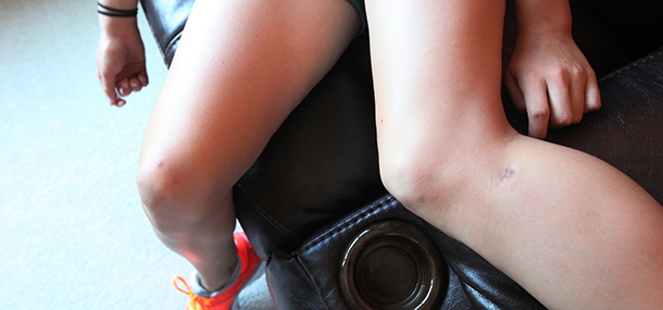
Pain in the front (anterior) part of the knee is a very common symptom in children. The knee is the largest joint in the body and where most growth occurs. The thigh bone (femur), upper shin bone (tibia), and knee cap (patella) are the major bones that make up the knee joint. They are connected to each other by ligaments and connected to the strong muscles of the thigh and calf by tendons.
Most children with anterior knee pain will not have a structural problem. Symptoms are often due to thigh muscle imbalance and/or overuse. Your child’s doctor will examine the knee and order x/rays to exclude structural abnormalities. The main treatment for this type of pain is activity modification and physical therapy with muscle strengthening/flexibility.
There are other symptoms such as swelling, locking, or instability that are more commonly associated with structural, traumatic, or inflammatory conditions. Your child’s doctor may want to further investigate with MR imaging.
Image: Arrows indicating Osgood-Schlatter disease
The large muscles in the front of the thigh responsible for extending the knee attach to the patella and tibia. These developing tendon attachments are subjected to a lot of physical stress in adolescence and can cause pain at the lower edge of the patella (Sinding/Larsen/Johansson disease) or at the upper edge of the tibia (Osgood/Schlatter disease). Doctors will often make this diagnosis without imaging, and treatment is conservative.
Image: Arrows indicating Osgood-Schlatter disease
The groove where the patella fits into the front of the femur or the patella itself may not be properly formed. This common condition results in abnormal movement of the knee as well as instability. It can also cause the knee to be more susceptible to dislocation, which is a condition of sudden worsening pain known as acute patellar dislocation. MRI is often used to identify the structural makeup of the knee and determine if an injury is present.
Anterior knee pain is a common, often chronic problem in children and adolescents and usually not due to a serious medical problem. Evaluation by a qualified physician/orthopedist and knee x-rays will typically diagnose the cause. MR imaging is reserved for those with more extensive symptoms or injury.
Contributed by Dr. Kathleen Emery and edited by Bessie Ganim (RT-NucMed).
