Vascular anomalies are disorders of blood vessels. Typically found in young children, they frequently present themselves as regions of skin discoloration and/or masses. They can generally be divided into two categories based on their cellular properties and behavior. The first group consists of true tumors or neoplasms, which may or may not be present at birth and grow out of proportion to the patient. These lesions are most commonly benign (harmless), with the classic example being the raised “bumpy strawberry” mark of an infantile hemangioma (or IH) on a young infant’s skin.
Even though it is very rare, some vascular tumors can be more aggressive or malignant (harmful) in particular cases. The infantile hemangioma has a very predictable life cycle as it is typically not present at birth, grows rapidly in the first few weeks to months of life, and then slowly resolves over several years. IHs rarely require treatment unless they are causing problems because of a specific critical location (e.g., around the eye or involving the airway), implication (as skin IHs found in certain numbers or patterns may allude to internal anomalies of greater significance), or superficial complication (such as ulceration, bleeding, and cosmetic deformity).
In contrast, vascular malformations are almost always present at birth as they represent regions of vessels that did not form properly while in the womb. However, they may not be noticed by a caregiver until later in childhood. The manifestations of these types of vascular anomalies can be quite variable as they can arise, in isolation or combination, from any of the types of vessels normally found in the body. Additionally, they may be small or large, focal or diffuse, and singular or multiple.
 Image: Ultrasound scan
Imaging may be employed in the setting of a vascular anomaly when its location, size, number, distribution, or an associated complication necessitates further assessment. Additionally, vascular anomalies may sometimes occur in the deeper tissues beneath the skin, in which case the characteristic (and frequently diagnostic) skin lesion may not be present. Such lesions will often undergo imaging in an attempt to establish the diagnosis and separate it from other masses, which may require different treatments (such as an inflammatory mass or a non-vascular tumor).
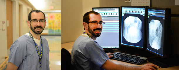
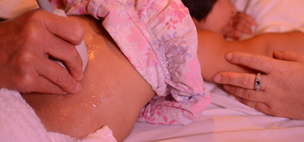
Ultrasound is typically used as the initial imaging approach to a touchable soft tissue mass in your child, as it requires no ionizing radiation or sedation and can yield high-quality images of the superficial soft tissues. It is particularly useful in obtaining information about the blood vessels in a mass, which can help establish the diagnosis of the vascular anomaly.
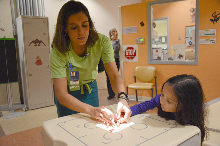
Another common imaging modality used in this setting is MRI. While typically requiring sedation for younger children, MRI uses no ionizing radiation and can evaluate some properties of a mass better than ultrasound. If the diagnosis remains unclear after these two studies, the mass may need to be sampled surgically to determine the appropriate treatment, if any.
 Image: MRI scan of the liver, showing innumerable round, well-circumscribed lesions
At Cincinnati Children’s, our Radiology Department works closely with a team of oncologists, surgeons, and pathologists who specialize in the diagnosis and treatment of vascular anomalies (known as the “Hemangioma and Vascular Malformation Center”). This group holds a weekly collaborative conference to discuss the details of each new and follow-up vascular anomaly patient in order to ensure that our center is providing your child and family with the best available care.
Contributed by Dr. Carl Merrow and edited by Tony Dandino (RT-MR).
Tony is an MRI Technologist at Cincinnati Children’s. Tony has been in his role for several years and serves as a Charge Tech, Quality Improvement Coach and Safety Coach for the MRI department. Tony has always known he wanted to work with children and in the medical field. Working at Cincinnati Children's has been the best of both worlds. Every day is something new and Tony can never wait to start the next adventure.