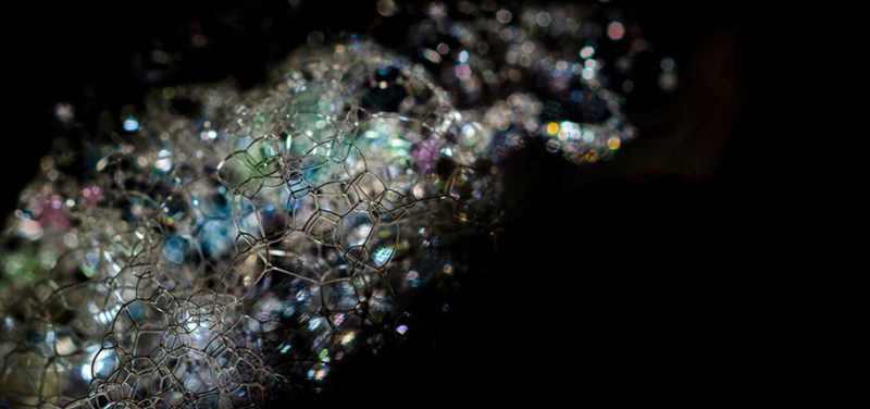
Featured Image: Only for display purposes only, not of actual microbubbles
Ultrasound is often the first choice for imaging children because it is not invasive, does not use radiation, does not require sedation, and is portable. At Cincinnati Children’s we use it a lot, everyday! Our Ultrasound Division is one of the busiest in the United States, performing over 33,000 studies in 2017!
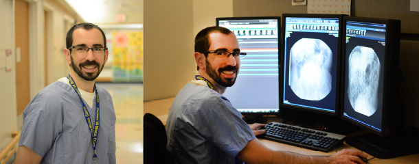
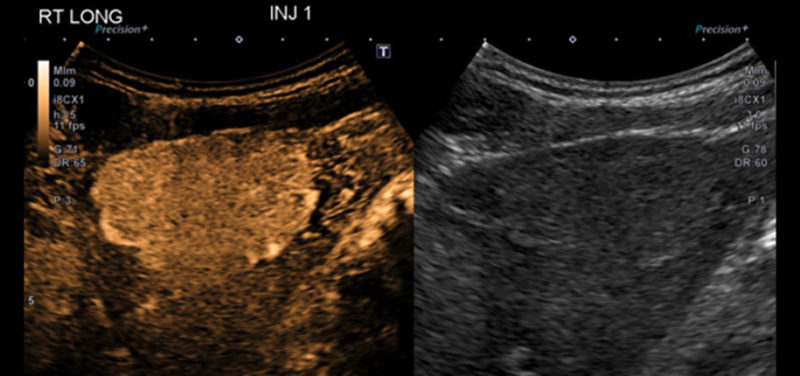
Even though ultrasound is great at demonstrating anatomy, it has a few limitations. In some areas of the body, particularly the liver, focal lesions can blend in with the surrounding normal tissue. These lesions can be very difficult to see with ultrasound and had to be further characterized with MRI or CT scans. Recently, the FDA approved the use of a microbubble contrast agent, Lumason™, which when injected intravenously perfuses organs and accentuates subtle differences in tissue that can make these lesions readily visible. In addition, the pattern of perfusion helps to further characterize these types of lesions, potentially making biopsy unnecessary. Using microbubble ultrasound contrast can eliminate the need for an MRI exam and answer clinical questions about focal liver lesions in patients who cannot safely have an MRI due to pacemakers or other metallic devices.
Cincinnati Children’s is currently offering contrast enhanced ultrasound exams to further characterize lesions in the liver and other parts of the body where perfusion characteristics help to narrow the diagnostic possibilities. We are also part of a multi-center trial evaluating the use of contrast during abdominal ultrasound screening of trauma patients. Preliminary studies prior to the multi-center trial showed very promising results. Additionally, we are exploring using contrast bubbles infused into the urinary bladder, to look for vesicoureteral reflux (VUR), similar to fluoroscopic VCUG and nuclear cystogram studies.
If contrast enhanced ultrasound sounds like a technique that may help you or your child, please discuss it with your physician or contact our ultrasound division for more information.
Contributed by Dr. Sara O’Hara and edited by Michelle Gramke, (ADV TECH_US).

Michelle is a Sonographer and has worked at Cincinnati Children’s for almost 26 years. She loves her job and loves working with kids. She is a lifetime Westsider (a person that lives on the west side of Cincinnati) and is married with 3 kids.