Radiation exposure from medical tests is a hot topic in the news. While all of us are exposed to small amounts of radiation daily from sources such as the sun, the food we eat, and the buildings we live in, parents are rightfully concerned about the amount of radiation their children receive.
At Cincinnati Children’s, we are committed to performing appropriate imaging tests that answer the questions asked by your child’s doctor. When possible, we use types of medical imaging that are free of ionizing radiation, such as ultrasound and magnetic resonance imaging (MRI). However, in many cases medical imaging using x-ray, fluoroscopy or computed tomography (CT) is the best way to evaluate your child’s illness.
In our Radiology Department, we are continually striving to provide imaging tests at the lowest possible radiation dose. We carefully follow the guidelines of the Image Gently© campaign (www.imagegently.org), which were created by the alliance for radiation safety in pediatric imaging. Also, there are many quality improvement projects in our department focused on decreasing radiation dose.
Related Article: Keeping Radiation Doses As Low As Possible for Your Child
For example, one of our cardiothoracic radiologists, a doctor who looks at images of the heart, lungs and airway, is working with one of our fellows to see if there is a better way to look at infants with a lung abnormality known as CPAM (congenital pulmonary airway malformation). A CPAM is an abnormality of lung development that can make it difficult for a baby to breathe when he or she is born. A CPAM can also rarely lead to cancer if it is not removed. As a result, surgery is usually necessary to remove the CPAM.
Before surgery, a CT scan is often requested to see the size and location of the CPAM and to see if there are any abnormal blood vessels flowing into or out of the CPAM. This CT scan provides important information to the surgeon, which allows for faster operations with fewer complications.
Traditionally, the CT scan done to look at the CPAM is designed to focus on the blood vessels using a technique called CTA (CT angiography). Our research team found that using a standard CT technique was able to answer the surgeon’s questions just as well as the CTA technique and at HALF the radiation dose! See if you notice any difference between the image obtained with the traditional CTA technique and the image obtained with the standard CT technique, as shown below.
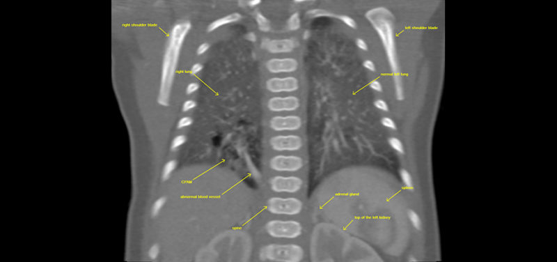 Image: CT coronal image right lower lobe CPAM with abnormal blood vessel.
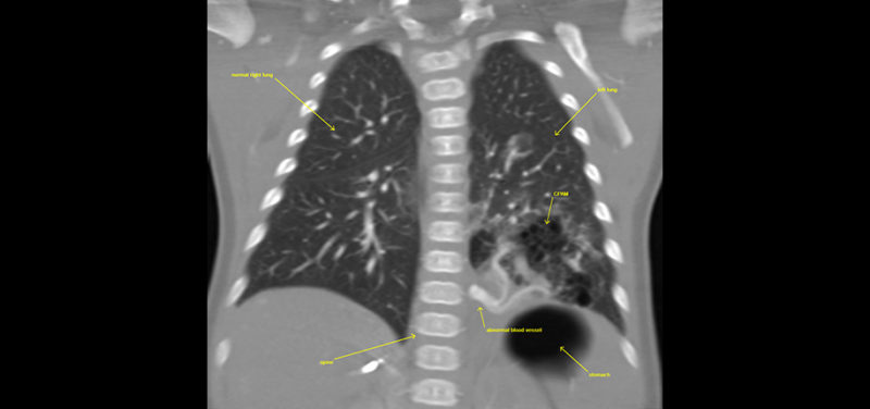 Image: CTA coronal image left lower lobe CPAM with abnormal vessel.
Our research team will share these findings with radiologists from around the world at the annual Society of Pediatric Radiology conference in Vancouver, BC in May.
Contributed by Karyn Ledbetter, MD and edited by Glenn Miñano, BFA.

Glenn Miñano is a media specialist in the Department of Radiology, providing graphic design, photography, printing, video services, and administration of the department’s online properties. His works have been published in several medical articles, such as the American Journal of Radiology and the American Institute of Ultrasound. He has been providing these services to the Radiology Department since 1996.