Most of us have been to a radiology department for an x-ray, ultrasound or even a CT or MRI. This kind of imaging provides a better look at what’s happening inside our bodies, whether it’s a simple broken bone, an inflamed appendix or a growing tumor. Radiologists are experts at looking at a variety of imaging to diagnosis diseases or conditions on a larger scale. As advances have been made in imaging, radiologists are able to now describe the type of tumor they see and even assign it a stage of progression that relates to prognosis or survival rate. Using this information, patients, families and the healthcare team can determine the next steps in the care plan.
 Figure 1. This image shows an MRI of hepatoblastoma in two separate children. Even though both children have the same type of tumor, their tumors look very different.
But what if the imaging could tell us more? What if radiologists could look at images and find more specific features or tiny details of a tumor to better predict how the tumor will behave in your child? What if our imaging could provide information that is so specific to your son or daughter that individualized therapy can be designed just for them? Radiomics and radiogenomics are growing fields in radiology that are trying to accomplish exactly this by trying to find relationships between tumor imaging, cell structure and tumor genetics. Several radiologists at Cincinnati Children’s are conducting research applying radiomics to pediatric diseases with encouraging results.
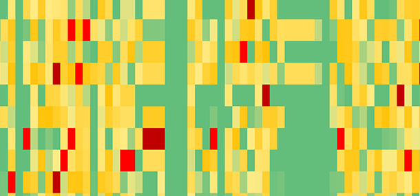
 Figure 2. The researchers reviewed each tumor for over 100 different characteristics in hopes of finding a pattern predicting the cells that make up the tumor. Areas on the map that are green are findings that are more likely to predict a specific type of tumor.
This new pediatric research that was recently presented at a scientific conference looked at MRI imaging already done on children with a rare liver tumor called hepatoblastoma. In initial studies, radiologists were able to identify several imaging features that correlated to specific tumor subtypes, a discovery that might impact clinical care since different subtypes of hepatoblastoma respond differently to treatment. While this research is just getting started, it will lead Cincinnati Children’s radiologists in new directions not only for hepatoblastoma but for other pediatric tumors. They will continue to search for predictors found in the imaging that directly relate to a more in-depth understanding of tumors and may lead to individualized treatments and therapies for children. In time, looking at imaging could be like gazing into a crystal ball, predicting the future and responding with all we’ve got!
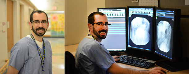
Story contributed by Dr. Alex Towbin, Dr. Andrew Trout, Dr. Matthew Plunk and edited by Catherine Leopard (Child Life).
Alex is a radiologist and the Neil D. Johnson Chair of Radiology Informatics. In this role, he helps to manage the information systems used by the Radiology department. Clinically, Alex is the Assistant Director of thoracoabdominal imaging. His research interests include liver disease, liver tumors, inflammatory bowel disease, and appendicitis.