There are many ways to evaluate the injured ankle in a child. Most often, the imaging evaluation begins with x-rays. This is a quick and relatively inexpensive method, and most often is all that is needed to make a diagnosis. However, radiographs can occasionally miss fractures as the images are two-dimensional; there can also be overlap of structures. Details of fractures or visualization of how extensive a fracture is can be challenging, particularly if the fracture is complex and extensive.
In cases where more information is needed, computed tomography (CT) can be very helpful to evaluate fractures. CT offers excellent anatomic detail and can show how displaced a fracture is or if it involves a joint. Two- and three-dimensional representations also can be made, which can be helpful for diagnosis. CT is more expensive and does require more radiation than radiographs. However, with the most updated scanners at Cincinnati Children’s, the radiation dose is kept very low. A CT scan also can be done with a cast in place with little effect on the images. Your doctor, usually an orthopedic surgeon or sports medicine physician, will take all these factors into account when ordering a CT scan for ankle injury.
Related Article: CT exams: What to expect from start to finish
Particularly at the ankle, it is important to make sure that the joint surfaces affected by fracture are smooth in order to prevent future arthritis and pain. Many fractures of the ankle are treated with casts, but a small number will need surgery to restore the normal anatomy. CT examinations can help the doctor plan the most appropriate treatment.
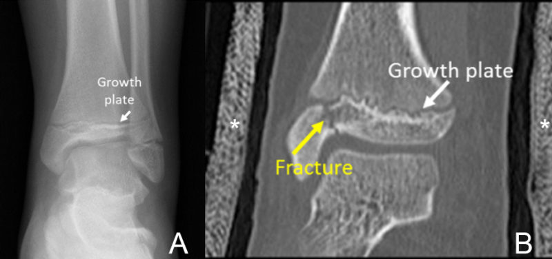
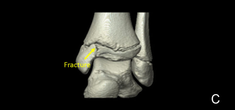
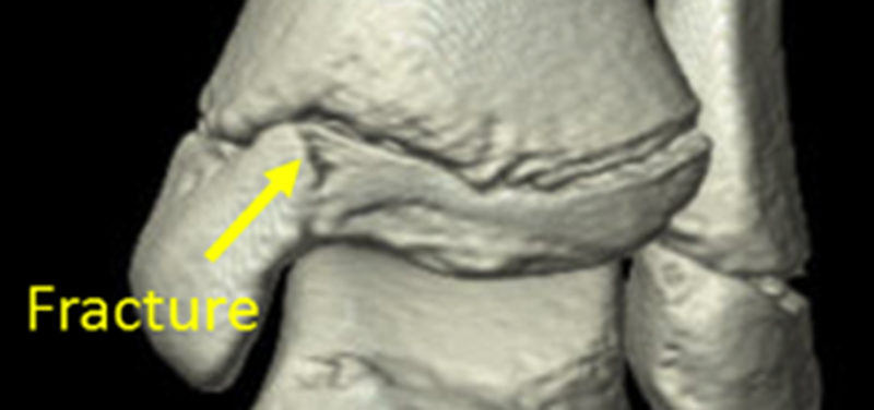
The radiograph (A) from a 12-year old girl who fell shows a normal growth plate. A fracture was suspected but barely visible. A CT scan was requested in order to better see the fracture. 2D (B) and 3D (C) images made from the CT scan taken with the ankle in a cast (asterisk) show the fracture line (yellow arrow) that goes to the ankle joint more easily. In this child, the joint surface is smooth and the fracture pieces are close together, so no surgery was necessary.
The ankle is one of the most common joints evaluated with CT after injury; however, many other bones and joints can be imaged also.
Contributed by Dr. Tal Laor and edited by Glenn Miñano, BFA.

Glenn Miñano is a media specialist in the Department of Radiology, providing graphic design, photography, printing, video services, and administration of the department’s online properties. His works have been published in several medical articles, such as the American Journal of Radiology and the American Institute of Ultrasound. He has been providing these services to the Radiology Department since 1996.