Functional MRI (fMRI) is a special type of MRI that uses changes in brightness on MRI images in relation to local changes in brain blood flow while performing a specific task to help localize brain function. fMRI has been used as a research tool for evaluating brain function for many years, and we now commonly use it to assess clinical patients who might undergo brain surgery for epilepsy, tumors, and other lesions.
The patterns of increased brightness (activation) can tell us, for example, where parts of the brain controlling hand movement are, where areas involved in understanding and language production are located, and where regions involved in vision are found. Using this information in addition to another special kind of MRI scan (diffusion tensor imaging or DTI) can also show us the connections between these important areas and other parts of the brain. Using the fMRI activation regions and the connections outlined by DTI, a road map for the surgeon can be created to help avoid any important areas during surgery.

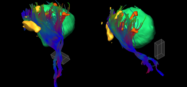
Diffusion Tensor Imaging (DTI)
fMRI exams are a little different than our usual MRI studies. To pinpoint areas important to language, vision, and movement, we have your child perform specific tasks while the MRI scan is being performed. These tasks are practiced with the patient prior to the exam, and are adjusted so that they can be performed based upon your child’s developmental stage and ability. Language tasks include listening to stories and thinking about words related to a presented object name. Visual tasks include watching a blinking checkerboard or looking at pictures and trying to remember them. Motor tasks may include moving fingers, feet or the tongue during the scan. Each patient gets a customized set of tasks based upon their ability and specific clinical issue. Given our experience working with children undergoing MRI and fMRI exams, we have been able to successfully perform fMRI exams on children as young as four years old!
As radiologists we often don’t get to interact as much as we would like with you and your families. With fMRI exams, however, this interaction is very important. When your child has an fMRI examination, one of the radiologists will assess and discuss the performance of the exam with you and your child on the day of the exam to ensure the best possible result. Currently, the patient must be awake and cooperative to perform the tasks needed for fMRI. In the near future, based upon research being done at Cincinnati Children’s and elsewhere, it may be possible to perform clinical fMRI exams while the patient is sedated, or while not doing any specific task. This type of fMRI, often called fMRI in the resting state, may help us obtain useful information on brain function on even younger children, those with more severe disabilities, and those requiring anesthesia to perform the exams.

Figure legend:
This is a 14-year-old girl with headaches. She had an enlarging tumor in the left frontal lobe (green). Prior to surgery the neurosurgeons wanted to know the best route into the tumor and how close areas controlling language were to the tumor. Areas active during production of verbs related to a presented noun are noted in orange. Areas active during tongue movement are in pink. Areas active during active speaking (in this case saying “ahhh”) are marked in blue. Note that the tumor is surrounded by very important language areas, some of them right on top of the tumor. After careful discussion of the risks and benefits of the procedure with the patient and family and using the fMRI and MRI scan as a guide, the surgeons were able to find a route in to the tumor missing the language areas for a complete removal. The patient had no speech problems after surgery.
Functional MRI is now a common tool for the assessment of children prior to brain surgery at Cincinnati Children’s. Our team of radiologists, technologists, and child life specialists’ work together to help children get the best result possible from the exam in a calm, child-centered environment.
Contributed by Dr. James Leach and edited by Tony Dandino (RT-MRI).
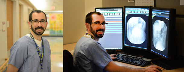
Tony is an MRI Technologist at Cincinnati Children’s. Tony has been in his role for several years and serves as a Charge Tech, Quality Improvement Coach and Safety Coach for the MRI department. Tony has always known he wanted to work with children and in the medical field. Working at Cincinnati Children's has been the best of both worlds. Every day is something new and Tony can never wait to start the next adventure.