When a girl is born with an anorectal malformation, she can sometimes have anomalies of her vagina, uterus, ovaries, kidneys or bladder. We’ll be focusing on vaginal anomalies, including imperforate hymen, partial agenesis of the vagina and complete agenesis of the vagina. These are congenital abnormalities and occur during embryological development, some of which may be diagnosed in infancy in the child’s life by fetal imaging or after birth by physical exam, ultrasound or MRI.
Many, however, may not be diagnosed until their female organs are under hormonal stimulation. The patient can have symptoms with severe pelvic pain or crampy pelvic pain. Radiology modalities of ultrasound and MRI can be helpful to determine type and any complications of these congenital anomalies. Although imaging is not necessary if the child’s history and physical exam lead the clinician to the appropriate clinical diagnosis, imaging can provide further information.
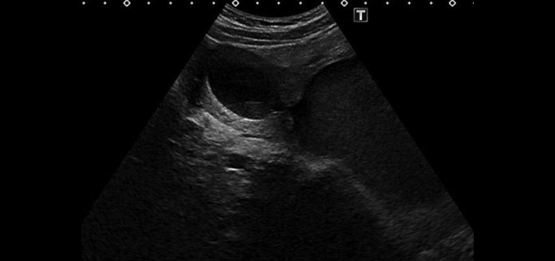
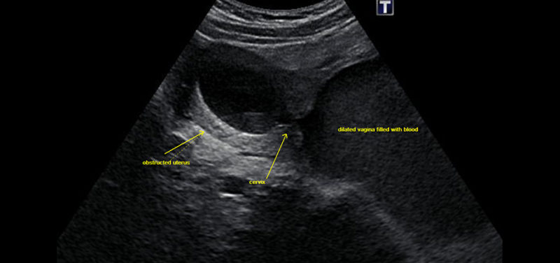 Image: Ultrasound scan of an obstructed uterus.
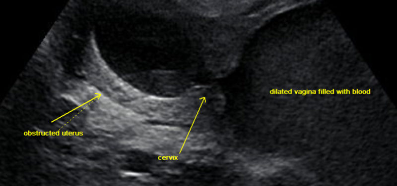 Image: Close-up of ultrasound image.
Ultrasound can demonstrate the dilated uterus and vagina, called a hematometrocolpos, which would be seen with vaginal obstruction from either imperforate hymen or partial agenesis of the vagina.
If your daughter has an anorectal abnormality or a kidney abnormality, she is more likely to have a uterus or vaginal abnormality, so be sure to keep this in mind and if she has pelvic pain. Further evaluation will be needed to determine any coexisting abnormalities of the female reproductive tract. Some girls will have normal periods from one side of the uterus while the other side of the uterus is obstructed.
Contributed by Dr. Kathy Helton-Skally and edited by Glenn Miñano, BFA.

Glenn Miñano is a media specialist in the Department of Radiology, providing graphic design, photography, printing, video services, and administration of the department’s online properties. His works have been published in several medical articles, such as the American Journal of Radiology and the American Institute of Ultrasound. He has been providing these services to the Radiology Department since 1996.