Subscribe today for more stories, tips, and updates.

Cincinnati Children's Blog
  • Subscribe
  • CincinnatiChildrens.org
  • Contact Us
  • Blog
  • Healthy Living
  • Rare and Complex Conditions
  • Safety and Prevention
  • Heart Conditions
  • Learning and Growing
  • Research and Discoveries
  • ?
  • Blog
  • Healthy Living
  • Rare and Complex Conditions
  • Safety and Prevention
  • Heart Conditions
  • Learning and Growing
  • Research and Discoveries
  • ?
  • Subscribe
  • CincinnatiChildrens.org
  • Contact Us
HOME/How We Do It/What’s Helium Got To Do With It?

What’s Helium Got To Do With It?

What’s Helium Got To Do With It?
October 11, 2021
0 Comments
By: Glenn Miñano

Most of us are familiar with helium: it makes party balloons float, and makes your voice sound funny if you breathe it in. Where does it come from? Most of it is found in natural gas reserves, so when natural gas is pumped out of the ground, helium is also collected. In the 1920s, the US government banned the export of helium and created the National Helium Reserve to store helium so that it could be used in military blimps. Thirty percent of the world’s stored helium remains in the reserve, but other countries have now created their own reserves. The United States and Qatar together produce about 25% of the world’s helium. 

MRI_Machine_DSC_2474_915x430

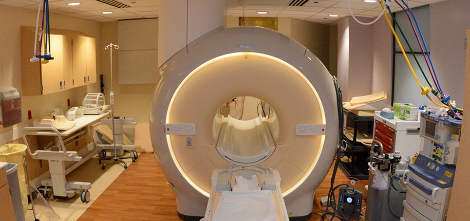
What does this have to do with radiology? Well, the single largest use of helium is as a cryogen in MRI machines. Cryogens are things used to maintain very low temperatures, like ice in a cooler. When helium is in liquid form, its temperature is more than 450 degrees below zero! The coils used in MRI machines have to be kept near this temperature in order to create the powerful magnetic field used to create images, so MRI machines have liquid helium surrounding the coils. If the helium goes from a liquid to a gas, the temperature quickly rises and the magnetic field goes away. Heating the helium so that it boils off like this is called “quenching” the MRI scanner, and it is done only if there is an emergency that requires turning off the magnetic field very quickly.  

When helium goes from a liquid to a gas, it takes up 750 times the space that it did as a liquid. If the helium boils off into the MRI scanner room, it will push all the oxygen out of the room and anybody inside the room will quickly lose consciousness. In order to prevent that from happening, MRI machines have vents that allow the helium to be expelled outside the building if a quench occurs. 

Recently, MRI designers have built MR scanners that use only a tiny amount of helium. If a quench happens, the helium gas stays inside the machine and doesn’t escape into the room at all, so there is no danger to the people in the room.  Our newest MRI scanners, located in the new critical care building and at our outpatient Kenwood site, have this design, using only 7 liters of helium as compared to the 1500 liters used in standard MR scanners. As our other MRI scanners get replaced, they will also use this new design, reducing the need for (and potential risk from) large amounts of helium. And leaving more for the rest of us to use in balloons. 


Jones template

Dr. Blaise Jones, author; Glenn Minano, BFA, editor; Meredith Towbin, copy editor 

Subscribe today for more stories, tips, and updates.

Related Articles

Radiologic Technologist Programs
<strong>MRI’s Role in Your Child’s Proton Therapy</strong> 
MRI’s Role in Your Child’s Proton Therapy 
<strong>Radiology’s Got Heart</strong>
Radiology’s Got Heart
Happy Saint Patric’s Day!
TAGS:
  • helium
  • radiology
Avatar photo

About the author: Glenn Miñano

Glenn Miñano is a media specialist in the Department of Radiology, providing graphic design, photography, printing, video services, and administration of the department’s online properties. His works have been published in several medical articles, such as the American Journal of Radiology and the American Institute of Ultrasound. He has been providing these services to the Radiology Department since 1996.

Write A Comment

Leave a Reply Cancel reply

Your email address will not be published. Required fields are marked *

No Comments Yet

Topics

  • Cancer and Blood Diseases
  • Child Development and Behavior
  • Childhood Obesity
  • Chronic Care Networks
  • Cincinnati Walks for Kids
  • Featured Faces
  • Fetal Conditions
  • Fitness and Nutrition
  • Food Allergies
  • Ghana 2012
  • Health Care Public Policy
  • Health Care Quality
  • Health Care Reform
  • Healthy Living
  • Heart Conditions
  • Hospital Operations
  • How We Do It
  • Learning and Growing
  • Meet The Team
  • Nurses Week 2012
  • Patient Family Experience
  • Patient Flow
  • Patient Safety
  • Patient Stories
  • Pin of the Week
  • Radiology
  • Rare and Complex Conditions
  • Research
  • Research and Discoveries
  • Safety and Prevention
  • Share Your Story
  • Technology
  • Uncategorized
  • Vaccines
  • Volunteerism
  • What Makes Us Different
  • What's New
See All Topics
See Less Topics

Subscribe

Never miss a post! Sign up to get new blog updates delivered to your email.

Popular Posts

  • 6 Questions to Ask When Your Child Complains of Chest Pain 2k views
  • Caring for the Caregivers of Children with Congenital Heart Disease  1.4k views
  • Stuttering In Young Kids: When To Be Concerned 877 views
  • Laryngomalacia: Is My Child’s Noisy Breathing Serious? 645 views
  • 4 Questions to Ask When Your Child Has Recurrent Croup 574 views
  • What’s the difference between a CAT-Scan and a CT-Scan? 500 views
About

The Cincinnati Children’s blog features thoughts and perspectives from our experts on all things pediatric health. This blog does not provide medical advice, diagnosis, or treatment. Learn More »

Contact Us
  • 3333 Burnet Avenue, Cincinnati, Ohio 45229-3026
  • 1-513-636-4200 | 1-800-344-2462
  • TTY: 1-513-636-4900
  • socialmedia@cchmc.org
Connect With Us
  • Subscribe
  • CincinnatiChildrens.org
  • Contact Us

© 2009-2022 Cincinnati Children’s Hospital Medical Center