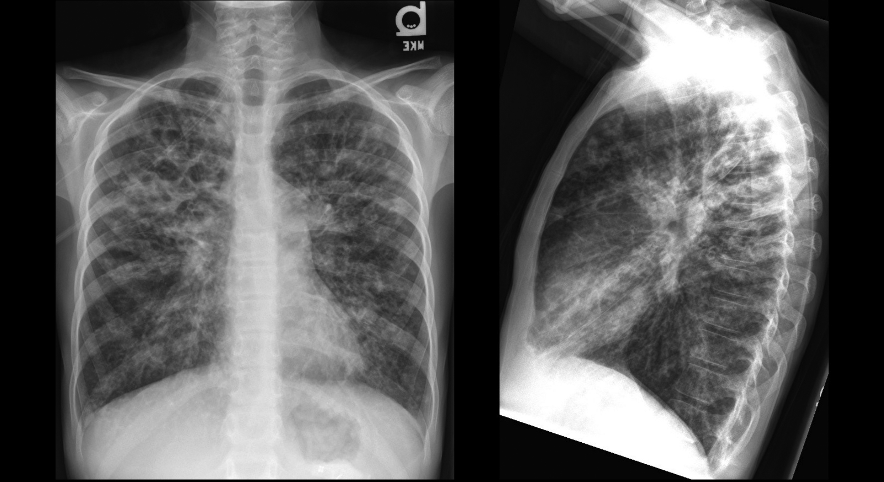
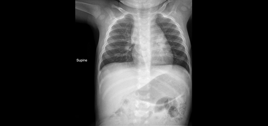
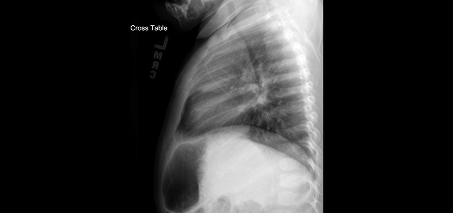
Sometimes, we are asked by parents why more than one view is needed for their child’s x-ray. Most of the time, the reason is because each image is a two-dimensional (2D) picture of a three-dimensional (3D) object (your child’s body). This means that many structures are superimposed upon each other. In other words, these structures appear to be on top of each other or blend in with each other. The second view (and sometimes more) is needed to separate out these structures for us to know where they exist in that 3rd dimension and more fully understand the anatomy.
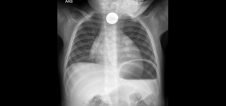
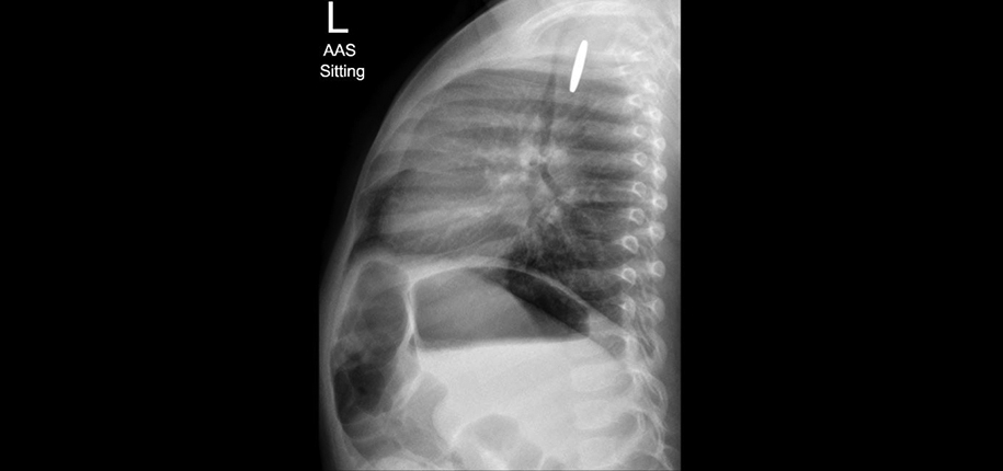
An example might be if your child swallowed a foreign body, such as a coin. If we see it in the upper chest, there may be question as to whether it lies in the trachea (airway) or the esophagus. Although we can usually tell by clinical presentation and the orientation of the object on the x-ray, there may be times when this is unclear. Also, if we see it in the upper abdomen, we may need another view to decide if it lies in the stomach or the intestines.
Another reason would be if something is showing up on the x-ray that may be an “artifact,” that is, it isn’t really there but is showing up on the x-ray, so we need another image to be sure that it is not an important finding. Sometimes, there may be external clothing or material that shows up on the image, which may be simulating an abnormality or it may be hiding an important finding. So, the extra image is needed to figure this out.
Additionally, we may need another image in order to see a structure more clearly. When a “large” picture is taken that includes many structures, it produces a less clear picture of each individual structure. So, when a radiologist sees a finding that needs to be clarified, he/she may ask for a “smaller” more detailed image of that structure. For example, when x-rays of the whole hand are taken, it may cause a small or subtle fracture of a bone in the finger to look indistinct or blurry. The radiologist may ask for a more detailed image of the finger to be sure whether it is a fracture or not.
A fourth reason may be to show change of a finding in time. For example, if a small or early pneumonia or other finding is seen on a chest x-ray, your child’s doctor may want to know if it worsens or improves over time.
There are other reasons why more images may be needed, but the above stated reasons are some of the more common ones. If you ever question why this is being done, you are welcome to ask one of our staff to clarify this for you.

Dr. Sunny Pitt, author; Glenn Miñano, BFA, editor; Meredith Towbin, copyeditor.
Glenn Miñano is a media specialist in the Department of Radiology, providing graphic design, photography, printing, video services, and administration of the department’s online properties. His works have been published in several medical articles, such as the American Journal of Radiology and the American Institute of Ultrasound. He has been providing these services to the Radiology Department since 1996.