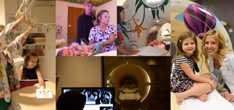
Cincinnati Children’s has a department that uses x-rays to take pictures of different parts of patients’ bodies. X-rays have been used in medicine since 1895 when the first picture of a hand was taken. Over the past 120 years, the use of x-rays in medicine has led to the development of other types of imaging and these are used everyday in the care of children. Nowadays, if you walk through any hospital’s “x-ray” department, you would see more than just “x-ray” signs. Instead you might see a variety of imaging types listed such as Ultrasound, CT, MRI and Nuclear Medicine.
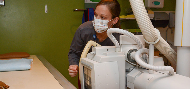

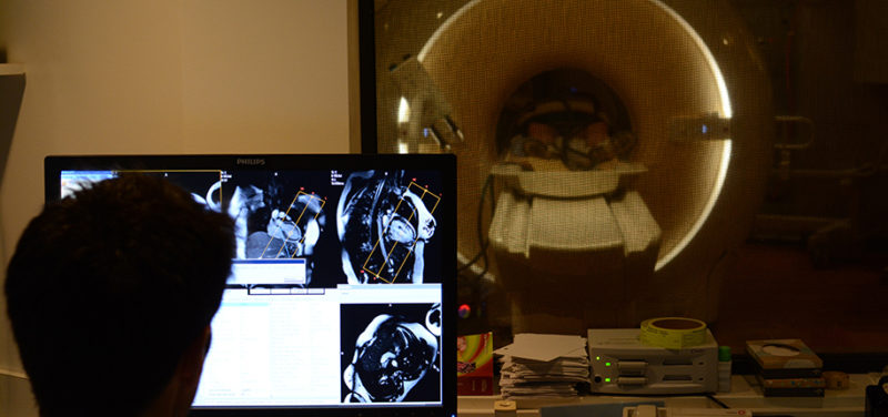
The different types of imaging that are found in our Radiology Department all have one thing in common: energy! Everything that happens in radiology uses energy of one kind or another. A common chest x-ray uses a higher energy form of electromagnetic radiation. This kind of energy can travel through things that ordinary light energy can’t. A CT scan also uses x-rays, but with the use of computer technology, it can turn a two-dimensional x-ray picture into a 3D image. Ultrasound and nuclear medicine utilize different kinds of energy for specialized medical imaging and treatments. Ultrasound uses sound wave energy to create images while nuclear medicine uses radioactive substances put into the body to create images and treat diseases. MRI scanners use radio wave energy (like the kind that lets you listen to music on your car stereo) and strong magnetic fields to create detailed images of the body.

Different kinds of energy are used for different kinds of imaging. Each type of energy creates unique pictures that show details that are important for diagnosis or treatment of a disease. Radiologists collaborate closely with specially trained radiology technologists and medical physicists to care for your child during imaging. This radiology team works carefully to control the amount of energy that is used to create each image, keeping your child safe while also getting the best quality pictures.
Our Department of Radiology and Medical Imaging Center at Cincinnati Children’s provides a wide range of care for pediatric patients with over 200,000 imaging procedures every year. Next time you come to our imaging department for an X-Ray, CT, MRI, Ultrasound, or some other type of “picture,” you can be assured that all of our staff are dedicated to obtaining highly accurate images while making those procedures safe and comfortable for your child.
Contributed by Keith J. Strauss, Ph.D and edited by Catherine Leopard, (Child Life Spec III).

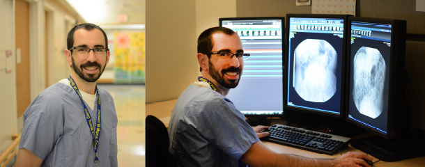
Alex is a radiologist and the Neil D. Johnson Chair of Radiology Informatics. In this role, he helps to manage the information systems used by the Radiology department. Clinically, Alex is the Assistant Director of thoracoabdominal imaging. His research interests include liver disease, liver tumors, inflammatory bowel disease, and appendicitis.