There are a lot of different types of imaging studies that your child’s provider may want to obtain to help answer a clinical question. One of the more unique studies is called fluoroscopy and it’s one you may have questions about.
Fluoroscopy is a type of study that uses x-rays, much like the typical x-ray you may get of your chest or knee, but it has the special ability to pulse these x-rays several times a second allowing for real-time imaging. What does this mean? Fluoroscopy is a like a live x-ray video of your child.
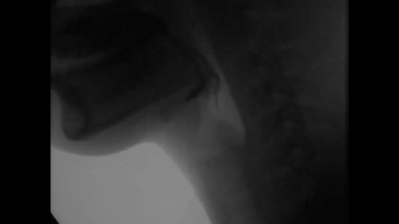
Video swallow study shows dynamic imaging of swallowing
It gives the radiologist and clinician information such as the anatomy of certain structures with quick results; it also provides dynamic information that may help answer why a certain symptom is present. It can be used to determine if a child is aspirating, to place tubes in a child with feeding difficulties, and to assess for abnormal things like reflux (from the stomach and the bladder). Additionally, this a radiology service that can help diagnose emergent conditions such as volvulus (bowel twisting on itself) and even treat emergent conditions such as intussusception (bowel telescoping into itself).
Studies ordered by your pediatrician such as an upper GI, esophagram, small bowel follow through, or video swallow study require your child to drink a material called contrast. The contrast is composed of a material that is radiodense, meaning that it shows up well on x-ray. This allows the radiologist to see structures like the stomach and small intestines better during the study. This contrast is inert and does not get absorbed by your child, eventually passing in his or her stool. While it might not be the best tasting thing in the world, our staff does their best to help sweeten it up with Kool-Aid flavors (go for fruit punch!).

While the fluoroscopy suites are relatively big rooms, by the time everyone is standing around the table, it can get quite crowded. With this large crowd of people, you or your child might be wondering who all of these people are or what their titles mean.
Fluoroscopy is a dynamic study. Often to see everything we need to see, your child will need to be constantly repositioned during the study (x-rays are 2D pictures and human bodies are 3D objects). Think of it like the roller coaster of radiology with lots of movement. The bed can even tilt head first and feet first, all the way to the upright position.
That is essentially the panel that receives the x-rays. The x-rays are generated from under the table and go through the table, your child, and then land on that box. After some convoluted physics, the image is generated and displayed on a screen.
X-rays are a form of radiation and the lead vest helps limit the amount of exposure vital organs receive. There are older blogs that go into more detail regarding radiation safety that I suggest you check out if you have more questions.
I hope this helps answer a little about an imaging modality that you may not know much about. Any questions you or your child may have will happily be answered at the time of your appointment by our wonderful staff at Cincinnati Children’s.

Dr. James Morris, author; Glenn Miñano, BFA, editor; Meredith Towbin, copyeditor
Glenn Miñano is a media specialist in the Department of Radiology, providing graphic design, photography, printing, video services, and administration of the department’s online properties. His works have been published in several medical articles, such as the American Journal of Radiology and the American Institute of Ultrasound. He has been providing these services to the Radiology Department since 1996.