This was a question I was recently asked by a precocious seven-year-old girl after I explained to her the procedure we were going to do that day: a functional MRI exam of her brain. I explained, “Well not really, but I can look at the blood flow in your brain while you think about words and move your fingers to tell what parts of the brain you are using.” My response was not entirely satisfactory. She looked at me quizzically then said, “Well, isn’t that reading your mind?” You be the judge.
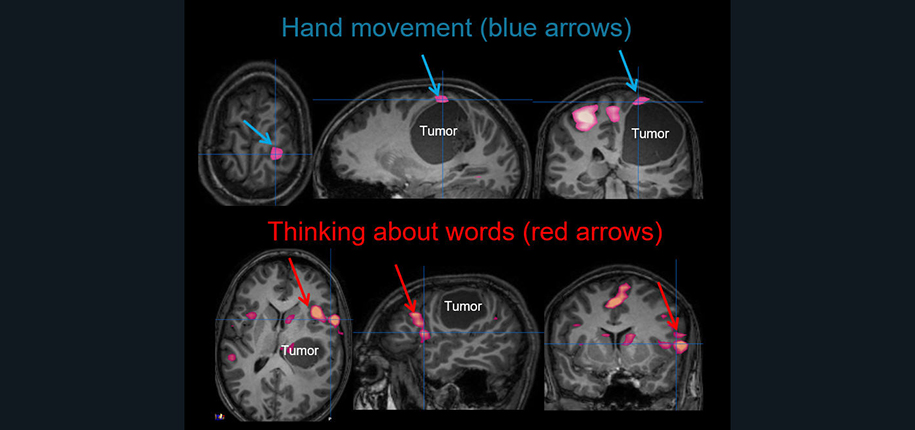
Functional MRI is a special type of MRI examination of the brain usually used in patients considering brain surgery for masses or for severe epilepsy. By using a special MRI technique and then processing the images on computers, we can identify areas of the brain involved in different tasks (language, movement, vision). During the fMRI examination, the patient will perform different tasks such as reading words and thinking about related action words, tapping their fingers, listening to stories, and/or watching images using a special sound system and TV screen made specifically for use in a MRI machine. I, Dr. James Leach, a radiologist with specific training and experience in imaging of the brain, performs the fMRI examination and guides patients through the study with the help of our tremendous Child Life staff and MRI technologists. After the examination is completed, I then process the information using a special computer and program to make images for the brain surgeon (Figure 1), Children older than six are typically the best candidates for fMRI examinations, although we have performed these exams on children as young as four years old.
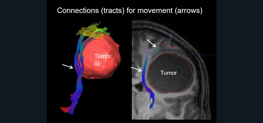
In addition, the functional MRI images are often used along with another special MRI technique called “diffusion tensor imaging,” or DTI. Whereas functional MRI looks at areas of brain function, DTI allows evaluation of the important brain connections or “wiring” connecting these specific areas with other regions of the brain or with the spinal cord. These white matter connections are often called “tracts.” A special computer program allows me to identify and outline these connections in a process called “tractography” (Figure 2). These are quite useful to the neurosurgeon as they will want to avoid these connections for maximum safety during surgery.
Cincinnati Childrens has been doing functional MRI exams and tractography for over 15 years and performs approximately two studies per week for surgical planning. Each study is planned and performed with one of our neuroradiologists specifically protocoling the exam for the child’s specific needs.
Contributed by Dr. James Leach and edited by Tony Dandino, (RT(MR))

Tony is an MRI Technologist at Cincinnati Children’s. Tony has been in his role for several years and serves as a Charge Tech, Quality Improvement Coach and Safety Coach for the MRI department. Tony has always known he wanted to work with children and in the medical field. Working at Cincinnati Children's has been the best of both worlds. Every day is something new and Tony can never wait to start the next adventure.