Radiating Hope: Radiology Department Blog

Search

Cardiac Magnetic Resonance Imaging (MRI): One-stop shopping

Post Date: April 7, 2021
Cardiac Magnetic Resonance Imaging (MRI): One-stop shopping

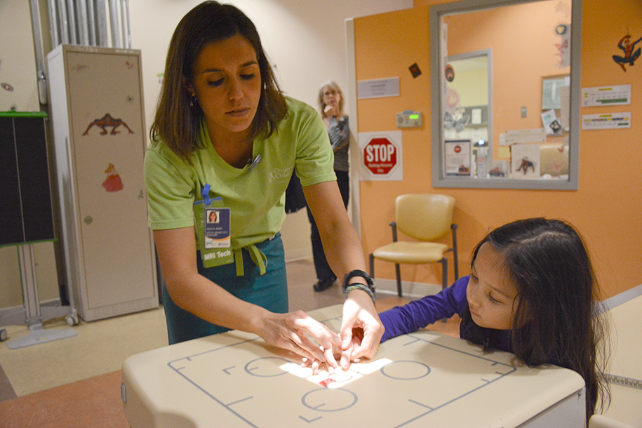
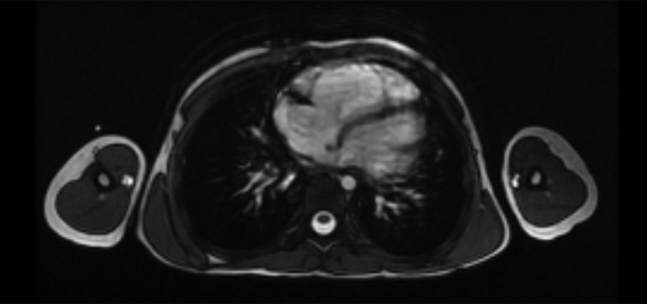
Cardiac or heart MRI is used to noninvasively image congenital heart defects and acquired diseases of heart muscles, chambers and vessels including coronary arteries. An MRI study is not painful and uses radio waves, not radiation. The best results for the majority of cardiac MRI techniques involve breath holding; however, the study can also be carried out during free breathing in patients who cannot hold their breath. Sedation may be necessary in small children.

Intravenous contrast agent is necessary to diagnose some diseases. However, patients should be questioned to determine the possibility of allergic reaction. Patients also need to be asked about the presence of metallic objects, implants, drug pumps, cardiac pacemaker, cochlear implants.

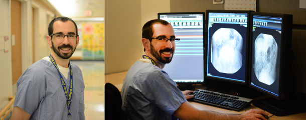
An average study takes about 45 minutes and images are evaluated by both a radiologist and cardiologist that specializes in cardiac imaging.

What information can be gathered by a cardiac MRI?

  • Structure of the heart, which is especially important in diagnosis and follow-up of congenital heart disease.
  • Function of the heart. Currently, cardiac MRI is regarded as the clinical gold standard imaging technique in measuring cardiac functions. Alternatively, noninvasive echocardiography is practical and widely available but has some limitations, including the inability to image deeper structures.
  • Measurement of blood flow within the vessels. MRI is considered the clinical gold standard imaging method in calculating blood flow velocity and output as well. This information helps to diagnose the narrowing of vessels and heart valves with valvular insufficiency.
  • Imaging of heart and other body vessels. This can be performed with or without intravenous contrast, and three dimensional images can be generated after an MRI study. This information gives surgeons a better overview of the heart and vessels before a surgery.
  • Virtual biopsy of the myocardium. With these unique characteristics, MRI can show certain tissue features without a real biopsy. This information is helpful to diagnose and follow up with myocardial infection and inflammation.

In summary, cardiac MRI can provide both structural, functional information and tissue characterization, allowing noninvasive, earlier and more accurate diagnosis of cardiac diseases without radiation.


Kocaoglu, Murat_template

Dr. Murat Kocaoglu, author; Glenn Miñano, BFA, editor; Meredith Towbin, copy editor

Avatar photo

About the author: Glenn Miñano

Glenn Miñano is a media specialist in the Department of Radiology, providing graphic design, photography, printing, video services, and administration of the department’s online properties. His works have been published in several medical articles, such as the American Journal of Radiology and the American Institute of Ultrasound. He has been providing these services to the Radiology Department since 1996.

About The Department

The Radiology Department at Cincinnati Children's is a leader in pediatric diagnostic imaging, radiology research, and radiation dose reduction.

Radiology Specialties