This last month, Cincinnati Children’s welcomed a new addition to its Nuclear Medicine Division in Radiology: a third-generation digital Positron Emission Tomography (PET) scanner. Not only is this scanner new to Cincinnati Children’s, but it’s new to the world! When the scanner was installed in November, it was the first clinical scanner of its type to be installed in a hospital anywhere in the world. The new scanner brings with it the most advanced engineering technology and computer software developed to date. With the new technology, we are able to scan our patients faster, at lower radiation doses, and with greater clarity.

The scanner is able to image faster because it has imaging components that are slightly longer and able to capture more radiation used to create the image. Historically, patients that received a PET examination may have been inside the scanner for up to an hour (depending on the height of the patient). The new scanner has already helped to reduce our overall imaging time drastically because of its new technology. Patients who come to Cincinnati Children’s for a PET examination are told not to eat beforehand (digesting food can prevent the best quality examination) and are not permitted to snack while in the hospital for several hours while they are prepared for the actual PET examination on the scanner. With the ability for the scanner to image faster, our patients will be able to get in and out of the hospital and back to their normal day faster. Additionally, for some of our younger patients, having a faster scanner should help reduce the need for sedation used to help these younger patients lie still during their examination.
PET imaging is subtly different than any other scanner in radiology that uses radiation to create an image. For PET imaging, we inject a small amount of radiation-based medicine that travels to the part of the body that the radiologist needs to see. Once in place, the medicine sends out radiation to the scanner to create the image. Because of the scanner’s ability to collect more radiation emitted from a patient faster, this allows us to use even lower amounts of radiation than we had already been using.
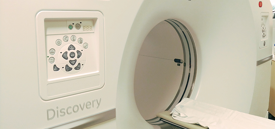
 Photo: Internal body of PET CT scanner
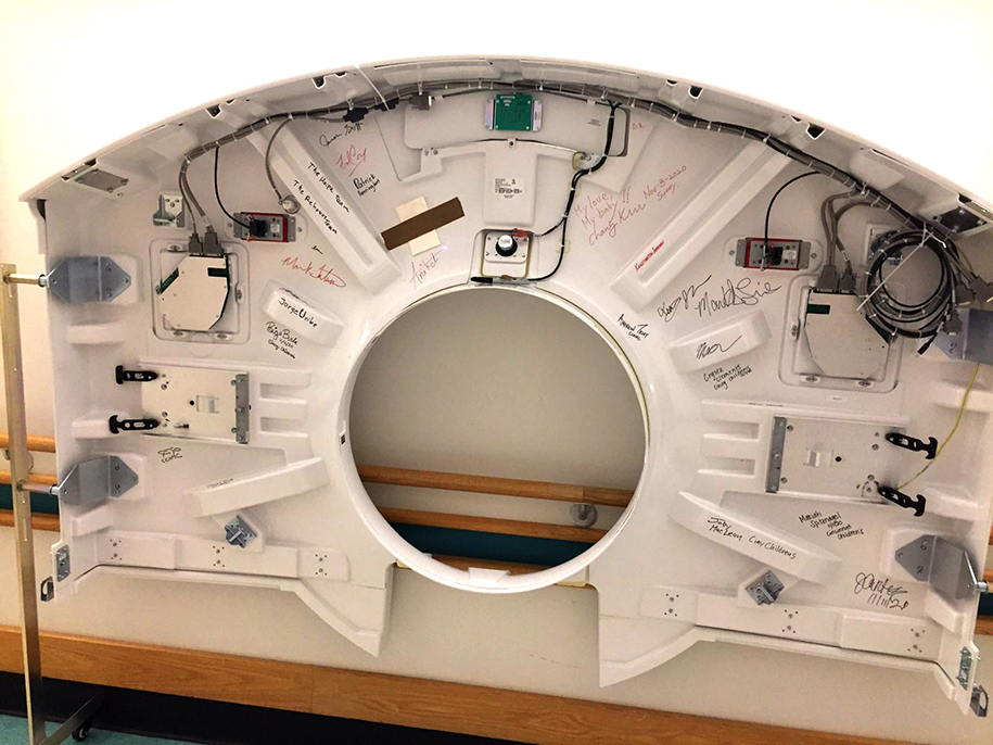 Photo: Internal body panel of PET CT scanner with signatures of the people involved with its creation and assembly
Finally, with the installation of the latest and greatest PET scanner, we have the ability to see more clearly. This new technology provides, for the first time, the ability to see even the smallest structures in the body that have been difficult to view in the past. With the ability to scan our patients faster, at lower radiation doses, and with more clarity, the new PET scanner gives the physicians at Cincinnati Children’s the most advanced tool to improve our patient healthcare and safety.


Samuel Brady, PhD, author; Glenn Miñano, BFA; Meredith Towbin, copyeditor; Joby MacLean, Rad Manager, internal photos contributions; Dr. Andrew Trout, PET CT scan image contribution
Glenn Miñano is a media specialist in the Department of Radiology, providing graphic design, photography, printing, video services, and administration of the department’s online properties. His works have been published in several medical articles, such as the American Journal of Radiology and the American Institute of Ultrasound. He has been providing these services to the Radiology Department since 1996.