Prenatal imaging with ultrasound is the standard of care for all pregnancies. However, when there is an abnormality, additional imaging with magnetic resonance imaging (MRI) is now more common.
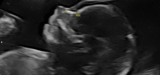
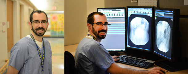
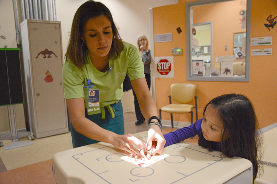
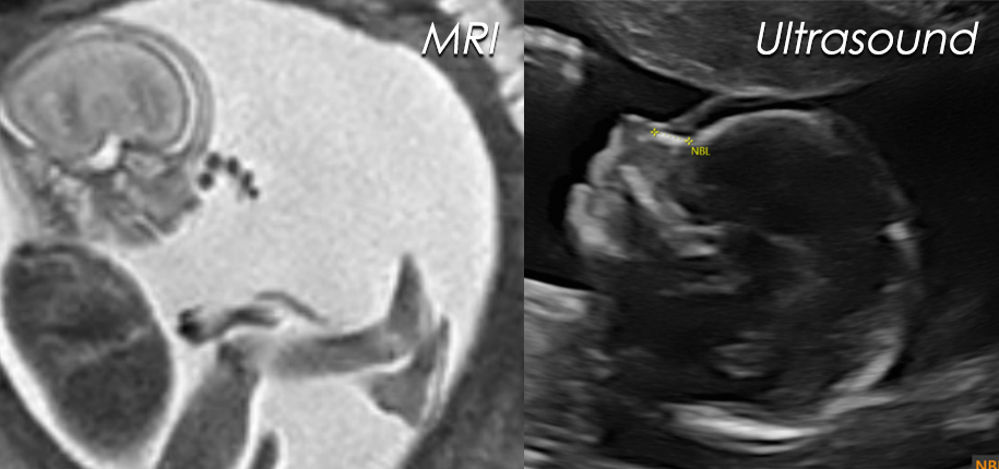
Why is this? Ultrasound is a test that utilizes sound waves. It provides amazing directed images of the fetus and allows us to reconstruct the images so we can see the bones and soft tissues, such as the fetal face. However, ultrasound and MRI together can tell us more. MRI is a test with radiofrequency waves and magnets, no radiation, and takes 40-45 minutes for a single baby. It provides excellent detail of the small structures in the growing fetus, especially the brain and lungs. With both ultrasound and MRI here at Cincinnati Children’s, the doctors now have a better understanding of the problems in the pregnancy.
By obtaining information with both prenatal ultrasound and MRI of abnormalities in growing babies, we are now able to direct care in the womb, at delivery and after birth. This has the potential to improve function and life expectancy for our smallest patients.
Dr. Beth M. Kline-Fath, author; Glenn Miñano, BFA, editor; Meredith Towbin, copyeditor

Glenn Miñano is a media specialist in the Department of Radiology, providing graphic design, photography, printing, video services, and administration of the department’s online properties. His works have been published in several medical articles, such as the American Journal of Radiology and the American Institute of Ultrasound. He has been providing these services to the Radiology Department since 1996.