Many children like to play barefoot, which means they face a greater risk of puncture wounds of the foot. These may go unrecognized at the time of injury and there may be little physical evidence of a puncture site. The only symptoms may be pain, local swelling, or signs of an inflammatory response.
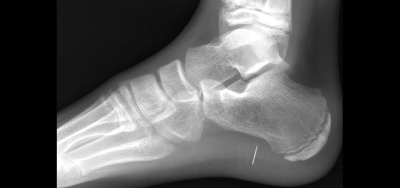 Image: Lateral x-ray of foot showing needle in heel
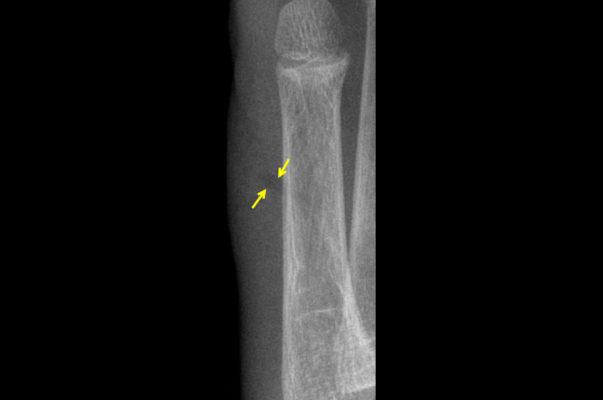 Image: Magnified x-ray of outside of foot with subtle darker linear structure in soft tissues (arrows) in child who stepped on a stick
When a child presents with foot pain, with or without the history of a suspected foreign body, x-rays of the foot are the usual first line of imaging. Not all foreign bodies will be seen on radiographs. Metallic objects like nails and pins are usually easily detected, but other common objects such as glass and wood/splinters are not easily seen. Depending on size and location, even some normally visible objects may be hard to visualize, particularly if not suspected or obscured by bone. Glass may have some metallic elements that allow visualization on x-ray. Wood fragments are notoriously difficult to see since they have no metallic components. However, there is usually some retained air within wood so it may appear as a more lucent structure (darker than the soft tissues) if x-rays are taken within a couple of days after the injury. As fluids around the object get absorbed, a wood foreign body will not be visible on x-ray.
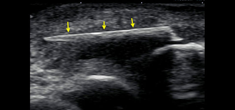 Image: Ultrasound image showing the bright linear structure of the retained wood fragment (arrows)
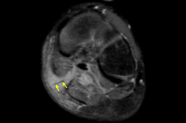 Image: MRI through mid-foot showing bright inflammatory changes in soft tissues with a dark linear structure that was found to be an unsuspected wood fragment (arrows)
If x-rays fail to identify a retained soft tissue foreign body and one is suspected, ultrasound is the next imaging modality of choice. Most objects will be readily identified as a bright (echogenic) structure. Often the surrounding inflammatory changes are easily shown. Ultrasound can be used to help guide removal of the structure. Sometimes an MRI is performed to assess a suspected soft tissue infection and an unexpected foreign body is detected. Wood material is the most common and will often appear as a dark signal structure with surrounding soft tissue, bright signal inflammatory tissue or abscess.
Most importantly, it is important to seek medical attention when there is suspicion of a puncture injury of the foot and/or possible retained foreign body as these can result in harmful infection of the soft tissues and/or bone. Foreign bodies typically require removal using aseptic technique with antibiotic therapy as determined by the caring physician or surgeon.
So watch your step!
Contributed by Dr. Kathleen Emery and edited by Michelle Gramke, (Adv Tech-US).

Michelle is a Sonographer and has worked at Cincinnati Children’s for almost 26 years. She loves her job and loves working with kids. She is a lifetime Westsider (a person that lives on the west side of Cincinnati) and is married with 3 kids.