We have come such a long way using ultrasound imaging to visualize internal organs of the body. As I interview some of the Cincinnati Children’s pioneers who were involved in the beginning stages of ultrasound imaging, I realize I am working with some brilliant individuals who have made a huge impact on ultrasound imaging. As a 40-year-old, I would think ultrasound has been around a lot longer than myself, but that is definitely not so. I feel honored to have trained and worked among some of the greatest: Dr. Diane Babcock, Tracey (Skelley) Adams, and Marsha (Chapman) Brody.

Photo: (lf-rt) Dr. Babcock, Marsha Brody and Tracey Adams
Today my children would say these individuals are the “Avengers” of ultrasound. The Avengers, the Earth’s mightiest heroes, the greatest super-team ever assembled, fought to be accepted and for the safe existence of Earth. The Avengers can be compared to these pioneers of ultrasound who fought so hard over the past 35 years to develop the technique of ultrasound and to train their sonographers at Cincinnati Children’s to become the best of the best. They fought for approval, and had to prove the reliability of ultrasound imaging.
Back in the day (1970s), Tracey and Marsha worked with Dr. LeQuesne to develop the ultrasound program. The first way to use ultrasound for imaging required the use of “water bath technology.” The body was imaged underwater in a large circular water tank. Thank goodness this wasn’t very popular in the United States because it looks quite uncomfortable. Fortunately for us, Dr. LeQuesne continued his ultrasound quest by training Dr. Diane Babcock, who is still currently working at Cincinnati Children’s today.

Dr. Babcock continues to research advances in ultrasound. In the late 1970s, Tracey and Marsha would scan baby heads together in the department: one person would hold the head and the other person would use the ultrasound probe to generate the ultrasound image. This would take up to two hours per exam for all the set-up, sedation, and scanning.

Eventually, Dr. Babcock obtained a portable machine that could travel to the Neonatal Intensive Care Unit for head ultrasounds. This decreased the scanning time because we did not have to transport the child to the Radiology Department. On the average, only six ultrasound exams were performed per day. As ultrasound evolved, more and more ultrasounds were ordered and surgeons accepted the reliability of the images. The neonatologists (baby doctors) accepted ultrasound for checking premature babies for hemorrhage (bleeds) into the brain and the neurosurgeons (Dr. Robert McLaurin) accepted ultrasound for diagnoses of hydrocephalus in spina bifida patients. Today, we do over 100 ultrasounds a day; each exam takes from 30-45 minutes. We do approximately 1800 ultrasound exams and over 200 portable exams each month.

Photo: Ultrasound scan of a head, (left) 1970s and (right) 2014.

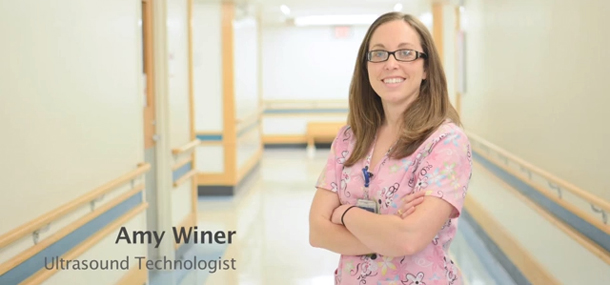
Photo: Modern ultrasound exam.
These three “Avengers” have helped develop ultrasound here at Cincinnati Children’s and they still have so much more to conquer. There are so many new ideas about ways to use ultrasound; improvements are always in the works. As of today, Dr. Babcock has passed the torch to Dr. Sara O’Hara, but she still remains active in her pursuit to make ultrasound the best it can be. Dr. O’Hara is now the director of Ultrasound and is carrying on the legacy of ultrasound. Her expertise and continued interest in ultrasound research has made our department one of the top sonography departments in the nation. Tracey Adams still actively manages and trains all of us sonographers, while Marsha Brody still has her mind set on defending the awesomeness of ultrasound but is currently working to raise three beautiful children.
As for me, I continue to learn something new and exciting everyday. I am so blessed to have been taught by such amazing individuals. I enjoy the excitement that Dr. Babcock brings to discussions of ultrasound. It is amazing to me that I work under the pioneers of ultrasound, and somehow they remain humble about their importance to the diagnostic advances of ultrasound. We are so blessed as employees and patients that they went the extra mile to create our future here at Cincinnati Children’s. This quest for the pursuit of happiness by our caregivers (surgeons and physicians) will continue to be their mission, and to advance the ultrasound imaging to help all mankind (our patients). As for me and my fellow sonographers, we have all helped in some small way to accomplish a greater good. Thank you for having a part in the success and evolution of ultrasound. The continued growth of ultrasound is what motivates me to continue my work at Cincinnati Children’s. We have become a part of a bigger universe.
Contributed by Stacie Sehlhorst (RT-RDMS) and edited bt Tony Dandino (RT-MRI).
Tony is an MRI Technologist at Cincinnati Children’s. Tony has been in his role for several years and serves as a Charge Tech, Quality Improvement Coach and Safety Coach for the MRI department. Tony has always known he wanted to work with children and in the medical field. Working at Cincinnati Children's has been the best of both worlds. Every day is something new and Tony can never wait to start the next adventure.