Subscribe today for more stories, tips, and updates.

Cincinnati Children's Blog
  • Subscribe
  • CincinnatiChildrens.org
  • Contact Us
  • Blog
  • Healthy Living
  • Rare and Complex Conditions
  • Safety and Prevention
  • Heart Conditions
  • Learning and Growing
  • Research and Discoveries
  • ?
  • Blog
  • Healthy Living
  • Rare and Complex Conditions
  • Safety and Prevention
  • Heart Conditions
  • Learning and Growing
  • Research and Discoveries
  • ?
  • Subscribe
  • CincinnatiChildrens.org
  • Contact Us
HOME/How We Do It/What are x-rays and how do they make a picture?

What are x-rays and how do they make a picture?

What are x-rays and how do they make a picture?
April 23, 2020
0 Comments
By: Glenn Miñano

There are several ways that radiologists make images of patients, but the oldest technique and one of the most widely known is the x-ray. The first image made using x-rays was produced in 1895 by Wilhelm Roentgen. But what exactly goes into creating those images?

We’re all used to using visible light to take pictures. In fact, since most cell phones have cameras, there’s a good chance you have a camera with you right now! When you visit our Radiology Department to have an x-ray exam, we use a special kind of camera to take a picture of the part of your body your doctor needs us to look at. To make it less confusing, radiologists call the beam of energy used to take these pictures an x-ray and the picture made using that beam of energy the radiograph.

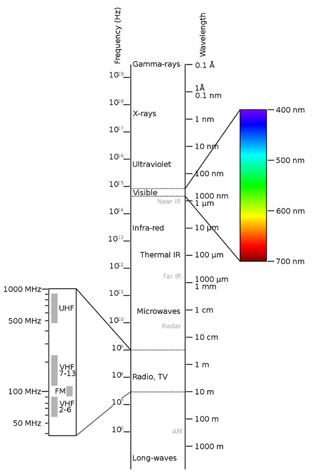
To understand what x-rays are, let’s talk about how they’re similar to light. Just like visible light travels from the sun through space to light up the earth, many other kinds of energy do the exact same thing. All these kinds of energy together make up the electromagnetic spectrum, and radiologists use several of the kinds of energy that we can’t see with our eyes to look inside the human body.

Electromagnetic spectrum chart


In radiology: 

  • Radio waves are used in MRI scanners 
  • X-rays are used in radiography 
  • Gamma rays are used in nuclear medicine 

We’re used to the ways visible light interacts with the world around us. Some things are transparent, or see-through, like a window. Other things are translucent, where some of the light passes through and some is blocked, like a lampshade. Other things are opaque and block light, like metal, wood, or a person. Just like visible light, x-rays are blocked more by some materials than by others. In the human body, bones block x-rays very well, while skin and muscle block them less. Parts of the body that contain air, like lungs, block x-rays even less. The amount of blocking, or attenuation, of the x-ray beam is related to density. Generally, the more dense an object, the better it blocks the x-ray beam. 

When an x-ray beam passes through a person, we can collect the energy on the other side to see what body parts blocked the beam. Just like the first visible light cameras used film to capture an image, the first x-ray cameras did, too. When the x-ray beam hit the film, it changed the color of the film, and that allowed the x-ray to be turned into a picture that could be seen with the eye. Just like most cameras we use these days are digital, though, so are many modern x-ray systems.   

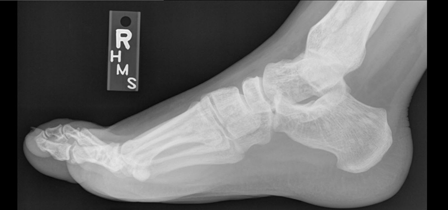
Pictures made using x-rays help doctors make many important diagnoses. They can help us see pneumonia in the lungs or a broken bone, just to name two examples. 

Being a radiologist doesn’t give you superpowers, but with a little help from science, we can use x-rays and other techniques to see inside the human body, which seems pretty close! 


Dr. Stacey Elangovan, author; Glenn Miñano, editor; Meredith Towbin, copyeditor.

Subscribe today for more stories, tips, and updates.

Related Articles

Arlene Schutte named as Radiology Family Recognition Award Winner
Happy Memorial Day
Happy Memorial Day
Rich Giordano Comes Back to Radiology to Fill Business Director Position
Jean Turner Minority Scholarship for Imaging Technology for 2023
TAGS:
  • make images of patients
  • radiology
  • x-rays
Avatar photo

About the author: Glenn Miñano

Glenn Miñano is a media specialist in the Department of Radiology, providing graphic design, photography, printing, video services, and administration of the department’s online properties. His works have been published in several medical articles, such as the American Journal of Radiology and the American Institute of Ultrasound. He has been providing these services to the Radiology Department since 1996.

Write A Comment

Leave a Reply Cancel reply

Your email address will not be published. Required fields are marked *

No Comments Yet

Topics

  • Cancer and Blood Diseases
  • Child Development and Behavior
  • Childhood Obesity
  • Chronic Care Networks
  • Cincinnati Walks for Kids
  • Featured Faces
  • Fetal Conditions
  • Fitness and Nutrition
  • Food Allergies
  • Ghana 2012
  • Health Care Public Policy
  • Health Care Quality
  • Health Care Reform
  • Healthy Living
  • Heart Conditions
  • Hospital Operations
  • How We Do It
  • Learning and Growing
  • Meet The Team
  • Nurses Week 2012
  • Patient Family Experience
  • Patient Flow
  • Patient Safety
  • Patient Stories
  • Pin of the Week
  • Radiology
  • Rare and Complex Conditions
  • Research
  • Research and Discoveries
  • Safety and Prevention
  • Share Your Story
  • Technology
  • Uncategorized
  • Vaccines
  • Volunteerism
  • What Makes Us Different
  • What's New
See All Topics
See Less Topics

Subscribe

Never miss a post! Sign up to get new blog updates delivered to your email.

Popular Posts

  • 6 Questions to Ask When Your Child Complains of Chest Pain 1.8k views
  • Stuttering In Young Kids: When To Be Concerned 887 views
  • 4 Questions to Ask When Your Child Has Recurrent Croup 617 views
  • Laryngomalacia: Is My Child’s Noisy Breathing Serious? 573 views
  • Is it safe for my child with braces to have an MRI? 441 views
  • 5 Reasons to Work with Child Life as an Adult with CHD 374 views
About

The Cincinnati Children’s blog features thoughts and perspectives from our experts on all things pediatric health. This blog does not provide medical advice, diagnosis, or treatment. Learn More »

Contact Us
  • 3333 Burnet Avenue, Cincinnati, Ohio 45229-3026
  • 1-513-636-4200 | 1-800-344-2462
  • TTY: 1-513-636-4900
  • socialmedia@cchmc.org
Connect With Us
  • Subscribe
  • CincinnatiChildrens.org
  • Contact Us

© 2009-2022 Cincinnati Children’s Hospital Medical Center