Most people have no idea what our imaging analysts’ duties consist of in the Post-Processing and Image Analysis Lab. The job is actually pretty elaborate! It’s time to take you on a little behind-the-scenes tour of what really happens.
 Image: “Sagittal Airway” image showing area covered in fly through video.
Video: “4D Virtual Endoscopic Fly Through” from pharynx down to the trachea. Patient history of subglottic stenosis and multiple airway reconstructions.
Employees must have training in Radiologic medical imaging. All of our imaging analysts previously worked or continue to work as CT or MRI technologists and maintain certification in either CT or MRI modalities. Employees in this department must exhibit proficiency in the utilization of electronic tools, paying close attention to details while creating and preparing imaging manipulation on virtually every part of the body. They work on a variety of software programs, acquiring extensive knowledge of human anatomy while working closely with our team of cardiologists, radiologists and the clinical MR physicist at Cincinnati Children’s. The work is diverse as are the talents and skills of the employees. One thing is for sure: no two days are alike.
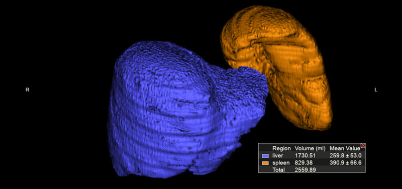
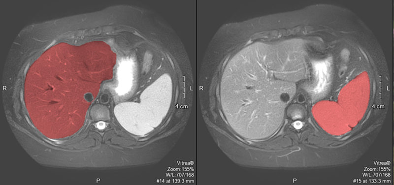 Images: “Liver/Spleen Volumes” performed in respect to rare genetic disorder where patients can experience enlarged liver/spleen.
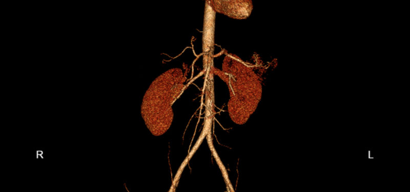
Just a few clinical examples of assessment through this department are MRI/CT cardiac function and analysis, MR hepatic & myocardial iron, MR elastography exams testing for liver stiffness, CT living donor candidates for kidney and liver transplants, MR fetal imaging, CT dynamic/static airway studies and MR brain perfusions. Providing this enhanced imaging manipulation and numerical data to the radiologists and physicians for review contributes to the overall improvement of patient care in the Radiology Department.
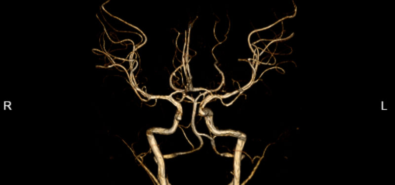 Image: “Circle of Willis” performed to detect blood vessel abnormalities.
Video: 3D rendering to demonstrate airway tree and lung periphery3D rendering to demonstrate airway tree and lung periphery.
Video: 3D batch to demonstrate relationship of airway to great vessels in a patient with cardiac abnormalities.
In addition to clinical work, the Post-Processing Department frequently participates in research projects. This research strengthens our current processes and continually improves outcomes for patients. A proud moment for several team members was taking part in the post-processing of the Peruvian child mummy (more than 500 years old) from the Cincinnati Museum Center scanned in CT at Cincinnati Children’s in 2015.
So remember, the next time you encounter “those pretty images” with your MRI or CT scan, they didn’t magically appear. A talented team of imaging analysts with 60+ years of experience at Cincinnati Children’s working behind the scenes in our Post-Processing and Image Analysis Lab completed that!
Contributed by Janice Steiner, BS, RT(R) (CT) and edited by Glenn Miñano, BFA.

Glenn Miñano is a media specialist in the Department of Radiology, providing graphic design, photography, printing, video services, and administration of the department’s online properties. His works have been published in several medical articles, such as the American Journal of Radiology and the American Institute of Ultrasound. He has been providing these services to the Radiology Department since 1996.