If your child has a history of inflammatory bowel disease, an enterography exam might be ordered by your physician. This exam looks at the small and large intestines as well as other organs in the abdomen and pelvis associated with inflammatory bowel disease. It is often ordered for patients with specific diseases including Crohn’s and ulcerative colitis.
An enterography exam can be performed using either MR or CT. It is generally preferred to use MR as the soft tissues are visualized better and there is the opportunity to perform imaging at different times after giving contrast (dye) through an IV. An MR exam is ideal for patients who are able to hold still for longer periods of time. CT scans are preferred for younger patients, patients who are unable to hold still as long, or if a patient cannot get an MR (due to having devices, implants, allergy to glucagon, or diabetes). While an MR scan is the preferred modality, a CT scan will provide optimized imaging as well.
For both MR and CT, your child will first need to not eat or drink for 4 hours prior to the scan. An IV needs to be started. This is one of the first things we do in case there is difficulty or issues so timing is not affected. Our team of technologists and child life specialists work with you and your child to make this as easy of a process as possible. In most instances, the IV is placed in the lower arm between the wrist and elbow, or in the hand.
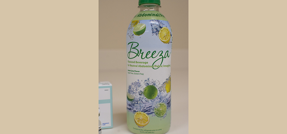
For the CT enterography, once the IV is placed, your child will start drinking the oral contrast (dye) called Breeza. This helps the small intestines be better seen on the imaging, with the goal being to fill the small intestines with Breeza at the time of imaging. The amount is based upon your child’s weight. Breeza is a more palatable drink compared to other drinks using contrast. It tastes like a flat sprite and can be flavored with sugar-free drink packets if requested. Your child needs to drink this continuously for 40 minutes. Before the last 15 minutes of drinking, your child will be brought into the scan room to lay on their right side while continuing the drink. Once the Breeza is finished, the scan is ready to start. Your child will lay on their back with their arms above their head. A second contrast (dye) is used through the IV, which works as a highlighter to visualize organs, blood vessels and other structures. When this contrast is administered, most patients feel a warm sensation in their neck, chest, and then abdomen. Some say they feel like they’ve wet their paints. All of these warm feelings are due to the opening of blood vessels, therefore increasing the blood flow. Those feelings usually go away shortly after the exam is complete.
When an MRI is indicated, there are few steps that need to happen once you arrive. The patient is brought back to the MRI changing room and changed into scrubs to remove all metal from clothing. The patient and family are then taken to an MRI prep room, and MRI screening is done. This is to make sure the patient is safe to enter the scan room. If a parent or guardian would like to accompany the child, they will be MRI “screened” as well. The magnetic field utilized to take MRI images is very strong, and certain implanted devices can be a danger to the patient or a family member who wants to go into the MRI.
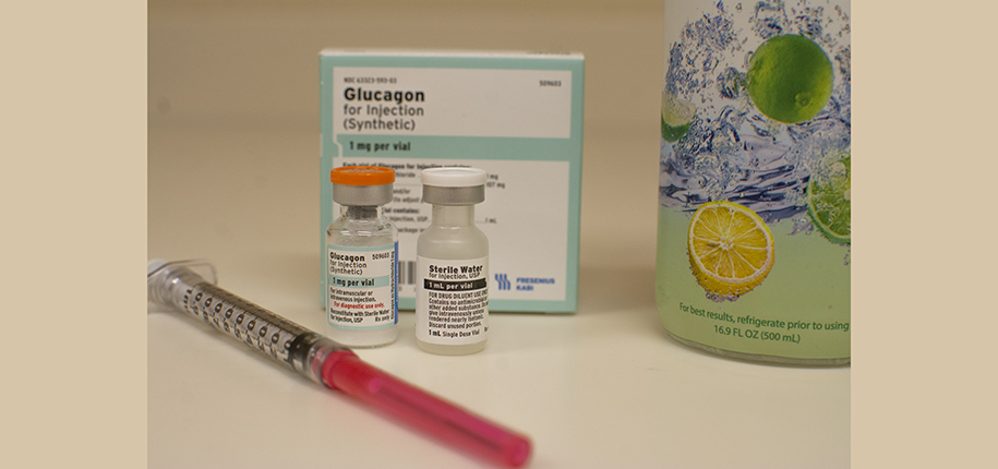
The next steps are very similar to the CT scan process. Your child will start by having an IV placed and drink the contrast continuously over a certain amount of time determined by the patient’s weight. In this time, the patient can pick out a movie or a type of music they would like to listen to. The MRI scanner makes loud construction like noises while it takes its pictures, and this helps protect the hearing as well as keep the patient entertained. Once in the scan room, the patient is given Glucagon through the IV, which slows down the movement of the intestines, allowing the contrast drink to stay where it needs to. Glucagon can cause some nausea and cramps, but it’s given very slowly to prevent those side effects from happening.
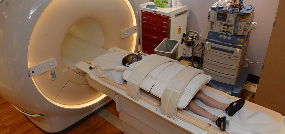
The patient is then set up with a special type of camera called a coil, which looks very similar to a turtle shell. This turtle shell is placed over the abdomen and allows us to get pictures in that region. Headphones and possibly movie goggles will be placed on the patient at this time. Early in the exam, another type of contrast called Dotarem is given by the IV. Unlike the drink contrast, this one will help show blood vessels and areas in the abdomen other than the intestines. This contrast should not make the patient feel different, other than a potential cold sensation up the arm and a smell or taste from the saline solution used to advance the contrast into the vein.
The MRI scan itself lasts anywhere from 40 minutes to over an hour. Some images are taken with breath-hold instructions, and some are taken as the patient breathes normally. The MRI technologist can communicate with the patient throughout the exam through the headphones used as hearing protection. A handheld squeeze ball is given to the patient in case of an emergency during the loud MRI image noises. A huge advantage to MRI is the variety of different images that can be acquired with great soft-tissue resolution. MRI uses physics to make different images with different “weightings” that are similar to a photo filter. This can help highlight abnormalities in the bowels that could be causing symptoms in a patient.
Breeza is used for both MR and CT exams. Some patients may experience loose stools after drinking it. There is no other special care required, and your child may resume normal activities after the scan unless your ordering provider advises you differently.
Results of these scans are released to the ordering physician within 48 hours. You can also view results and imaging using MyChart.
Collaboration with Dr. Rama Ayyala, Associate Professor of Radiology, Associate Chief, Culture, Quality, and Safety Division Director, Thoracoabdominal Imaging
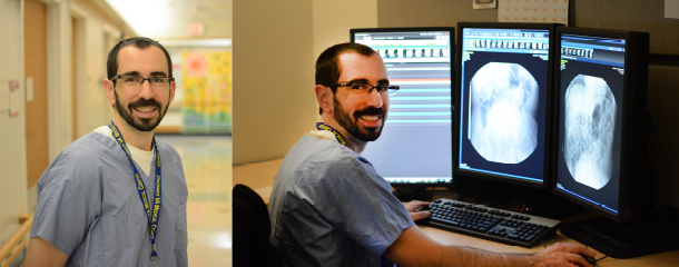
Jessica is a CT Tech who works primarily at Base and floats to Liberty CT. Her entire professional career of nearly 22 years has been at CCHMC in the Radiology Department. It was during her clinical rotation in x-ray school where she learned of her passion to work with children. In her free time, she and her husband enjoy cooking together and traveling. She has 2 adult stepsons and 2 dogs.
Erin McNamara is an MRI Technologist at Cincinnati Children's Burnet Campus. She’s also certified in nuclear medicine! Erin loves the pace of radiology here at Cincinnati Children’s, and how their job is always advancing and changing! 2023 she’s coming up on her two years at the hospital. It has been her dream job since middle school to work here, and it has not disappointed her. She likes adventuring around the tri-state area in her free time, watching sports, skiing, and hanging out with her cats and boyfriend.
Meredith Towbin is a freelance copy editor and writer. She has copyedited the Department of Radiology’s blog since it launched. She also works as a copy editor for the home improvement website BobVila.com. Her writing has been featured on HuffPost as well as other writing sites.