Imagine taking a fantastic voyage deep into your own eye. As you get smaller and smaller, the world around you becomes surreal.
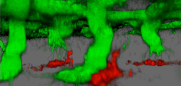
This is the developing retina of a mouse, as seen under a powerful confocal microscope. The green structures depict blood vessels reaching out to form new branches. The red objects are myeloid cells that act as cellular traffic cops guiding the process.
This image was provided by Richard Lang, PhD, director of the Visual Systems Group at Cincinnati Children’s. Lang is studying how blood vessels form in the retina to learn more about a common cause of blindness called macular degeneration. Surprisingly, his findings also may offer insights into controlling cancer.
Lang describes the molecular processes at work in a scientific paper published May 29, 2011 in the journal Nature.
If a drug can be developed that can slow down blood vessel formation, it could reduce the risk of macular degeneration, which is caused by out-of-control blood vessels damaging the retina. Slowing the process also could help fight cancer by cutting off the blood supply that tumors need to grow.


Tim this is so fascinating, the developing retina of a mouse under the microscope looks so odd yet intriguing! Let’s hope Richard Lang continues to work on this and is successful in reducing macular degeneration! 🙂 Thanks your friends at Busam Nissan.